• 文献检索
  • 文档翻译
  • 深度研究
  • 学术资讯
  • Suppr Zotero 插件Zotero 插件
  • 邀请有礼
  • 套餐&价格
  • 历史记录
应用&插件
Suppr Zotero 插件Zotero 插件浏览器插件Mac 客户端Windows 客户端微信小程序
定价
高级版会员购买积分包购买API积分包
服务
文献检索文档翻译深度研究API 文档MCP 服务
关于我们
关于 Suppr公司介绍联系我们用户协议隐私条款
关注我们

Suppr 超能文献

核心技术专利:CN118964589B侵权必究
粤ICP备2023148730 号-1Suppr @ 2026

文献检索

告别复杂PubMed语法,用中文像聊天一样搜索,搜遍4000万医学文献。AI智能推荐,让科研检索更轻松。

立即免费搜索

文件翻译

保留排版,准确专业,支持PDF/Word/PPT等文件格式,支持 12+语言互译。

免费翻译文档

深度研究

AI帮你快速写综述,25分钟生成高质量综述,智能提取关键信息,辅助科研写作。

立即免费体验

麻醉诱导意识丧失期间全球信息交换减少。

Global reduction of information exchange during anesthetic-induced unconsciousness.

机构信息

The Huck Institutes of Life Sciences, The Pennsylvania State University, University Park, PA, 16802, USA.

Department of Biomedical Engineering, The Pennsylvania State University, W-341 Millennium Science Complex, University Park, PA, 16802, USA.

出版信息

Brain Struct Funct. 2017 Sep;222(7):3205-3216. doi: 10.1007/s00429-017-1396-0. Epub 2017 Mar 13.

DOI:10.1007/s00429-017-1396-0
PMID:28289883
原文链接:https://pmc.ncbi.nlm.nih.gov/articles/PMC5597454/
Abstract

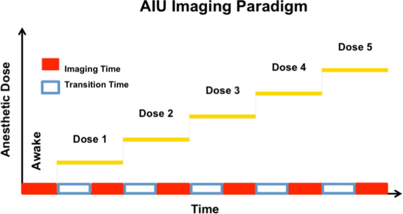
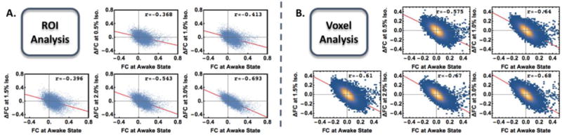
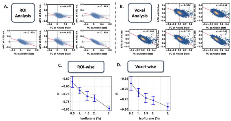
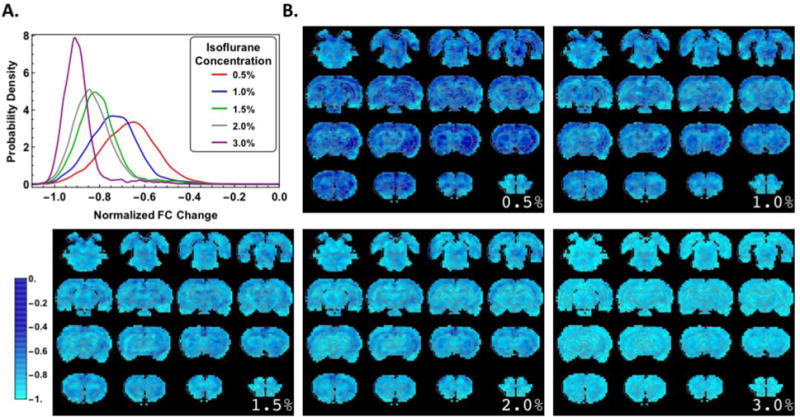
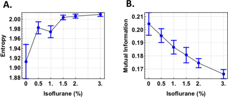
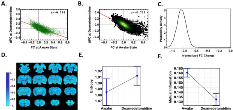
During anesthetic-induced unconsciousness (AIU), the brain undergoes a dramatic change in its capacity to exchange information between regions. However, the spatial distribution of information exchange loss/gain across the entire brain remains elusive. In the present study, we acquired and analyzed resting-state functional magnetic resonance imaging (rsfMRI) data in rats during wakefulness and graded levels of consciousness induced by incrementally increasing the concentration of isoflurane. We found that, regardless of spatial scale, the functional connectivity (FC) change (i.e., ∆FC) was proportionally dependent on the FC strength at the awake state across all connections. This dependency became stronger at higher doses of isoflurane. In addition, the relative FC change at each anesthetized condition (i.e., ∆FC normalized to the corresponding FC strength at the awake state) was exclusively negative across the whole brain, indicating a global loss of meaningful information exchange between brain regions during AIU. To further support this notion, we showed that during unconsciousness, the entropy of rsfMRI signal increased to a value comparable to random noise while the mutual information decreased appreciably. Importantly, consistent results were obtained when unconsciousness was induced by dexmedetomidine, an anesthetic agent with a distinct molecular action than isoflurane. This result indicates that the observed global reduction in information exchange may be agent invariant. Taken together, these findings provide compelling neuroimaging evidence suggesting that the brain undergoes a widespread disruption in the exchange of meaningful information during AIU and that this phenomenon may represent a common system-level neural mechanism of AIU.

摘要

在麻醉诱导的无意识(AIU)期间,大脑在区域之间交换信息的能力发生了巨大变化。然而,整个大脑中信息交换损失/增益的空间分布仍然难以捉摸。在本研究中,我们在清醒状态和递增异氟醚浓度诱导的不同意识水平下获取并分析了大鼠的静息态功能磁共振成像(rsfMRI)数据。我们发现,无论空间尺度如何,功能连接(FC)变化(即 ∆FC)与所有连接在清醒状态下的 FC 强度成正比。这种依赖性在异氟醚剂量更高时更强。此外,在每个麻醉状态下的相对 FC 变化(即 ∆FC 相对于清醒状态下的相应 FC 强度归一化)在整个大脑中均为负,表明在 AIU 期间大脑区域之间的有意义信息交换普遍丧失。为了进一步支持这一观点,我们表明在无意识状态下,rsfMRI 信号的熵增加到与随机噪声相当的值,而互信息则显著降低。重要的是,当使用具有与异氟醚不同分子作用的麻醉剂右美托咪定时,也获得了一致的结果。这一结果表明,观察到的信息交换总体减少可能是药物不变的。总之,这些发现提供了令人信服的神经影像学证据,表明大脑在 AIU 期间经历了有意义信息交换的广泛破坏,并且这种现象可能代表 AIU 的共同系统级神经机制。

https://cdn.ncbi.nlm.nih.gov/pmc/blobs/c5f1/5597454/6ed871cb02f7/nihms859921f9.jpg
https://cdn.ncbi.nlm.nih.gov/pmc/blobs/c5f1/5597454/8f8132585776/nihms859921f1.jpg
https://cdn.ncbi.nlm.nih.gov/pmc/blobs/c5f1/5597454/3f569c6552fa/nihms859921f2.jpg
https://cdn.ncbi.nlm.nih.gov/pmc/blobs/c5f1/5597454/7b69fc9694f2/nihms859921f3.jpg
https://cdn.ncbi.nlm.nih.gov/pmc/blobs/c5f1/5597454/341b18631659/nihms859921f4.jpg
https://cdn.ncbi.nlm.nih.gov/pmc/blobs/c5f1/5597454/428137e8b8f1/nihms859921f5.jpg
https://cdn.ncbi.nlm.nih.gov/pmc/blobs/c5f1/5597454/08d18c3b151a/nihms859921f6.jpg
https://cdn.ncbi.nlm.nih.gov/pmc/blobs/c5f1/5597454/923914e25c01/nihms859921f7.jpg
https://cdn.ncbi.nlm.nih.gov/pmc/blobs/c5f1/5597454/83d866053b4e/nihms859921f8.jpg
https://cdn.ncbi.nlm.nih.gov/pmc/blobs/c5f1/5597454/6ed871cb02f7/nihms859921f9.jpg

相似文献

1
Global reduction of information exchange during anesthetic-induced unconsciousness.麻醉诱导意识丧失期间全球信息交换减少。
Brain Struct Funct. 2017 Sep;222(7):3205-3216. doi: 10.1007/s00429-017-1396-0. Epub 2017 Mar 13.
2
Dynamic Connectivity Patterns in Conscious and Unconscious Brain.有意识和无意识大脑中的动态连接模式
Brain Connect. 2017 Feb;7(1):1-12. doi: 10.1089/brain.2016.0464. Epub 2016 Dec 15.
3
BOLD temporal variability differentiates wakefulness from anesthesia-induced unconsciousness.血氧水平依赖性功能磁共振成像的时间变异性可区分清醒状态与麻醉诱导的无意识状态。
J Neurophysiol. 2018 Mar 1;119(3):834-848. doi: 10.1152/jn.00714.2017. Epub 2017 Dec 6.
4
Simultaneous electroencephalographic and functional magnetic resonance imaging indicate impaired cortical top-down processing in association with anesthetic-induced unconsciousness.同时进行的脑电图和功能磁共振成像表明,与麻醉诱导的无意识相关,皮质顶叶自上而下的处理受到损害。
Anesthesiology. 2013 Nov;119(5):1031-42. doi: 10.1097/ALN.0b013e3182a7ca92.
5
Isoflurane induces dose-dependent alterations in the cortical connectivity profiles and dynamic properties of the brain's functional architecture.异氟烷会引起大脑功能结构的皮质连接图谱和动态特性发生剂量依赖性改变。
Hum Brain Mapp. 2014 Dec;35(12):5754-75. doi: 10.1002/hbm.22583. Epub 2014 Jul 12.
6
Impaired thalamocortical connectivity in humans during general-anesthetic-induced unconsciousness.全身麻醉诱导昏迷期间人类丘脑皮质连接受损。
Neuroimage. 2003 Jun;19(2 Pt 1):402-11. doi: 10.1016/s1053-8119(03)00103-4.
7
Disrupted neural variability during propofol-induced sedation and unconsciousness.异丙酚诱导镇静和意识丧失期间神经活动可变性的改变。
Hum Brain Mapp. 2018 Nov;39(11):4533-4544. doi: 10.1002/hbm.24304. Epub 2018 Jul 4.
8
Neural Correlates of Sevoflurane-induced Unconsciousness Identified by Simultaneous Functional Magnetic Resonance Imaging and Electroencephalography.通过同步功能磁共振成像和脑电图识别七氟醚诱导昏迷的神经关联
Anesthesiology. 2016 Nov;125(5):861-872. doi: 10.1097/ALN.0000000000001322.
9
Effects of isoflurane anesthesia on resting-state fMRI signals and functional connectivity within primary somatosensory cortex of monkeys.异氟烷麻醉对猴子初级体感皮层静息态功能磁共振成像信号及功能连接的影响。
Brain Behav. 2016 Oct 11;6(12):e00591. doi: 10.1002/brb3.591. eCollection 2016 Dec.
10
Common functional mechanisms underlying dynamic brain network changes across five general anesthetics: A rat fMRI study.五种全身麻醉药作用下动态脑网络变化的共同功能机制:一项大鼠功能磁共振成像研究
CNS Neurosci Ther. 2024 Jul;30(7):e14866. doi: 10.1111/cns.14866.

引用本文的文献

1
A novel integration of brain structural and functional connectivity for identifying traumatic brain injury induced perturbations.一种用于识别创伤性脑损伤所致扰动的脑结构与功能连接性的新型整合方法。
J Neurosci Methods. 2025 Jul;419:110459. doi: 10.1016/j.jneumeth.2025.110459. Epub 2025 Apr 22.
2
EiDA: A lossless approach for dynamic functional connectivity; application to fMRI data of a model of ageing.EiDA:一种用于动态功能连接性的无损方法;应用于衰老模型的功能磁共振成像数据。
Imaging Neurosci (Camb). 2024 Mar 22;2:1-22. doi: 10.1162/imag_a_00113. eCollection 2024 Mar 1.
3
Sequential Deactivation Across the Hippocampus-Thalamus-mPFC Pathway During Loss of Consciousness.

本文引用的文献

1
Dynamic Connectivity Patterns in Conscious and Unconscious Brain.有意识和无意识大脑中的动态连接模式
Brain Connect. 2017 Feb;7(1):1-12. doi: 10.1089/brain.2016.0464. Epub 2016 Dec 15.
2
Disruption of corticocortical information transfer during ketamine anesthesia in the primate brain.灵长类动物大脑在氯胺酮麻醉期间皮质-皮质信息传递的中断。
Neuroimage. 2016 Jul 1;134:459-465. doi: 10.1016/j.neuroimage.2016.04.039. Epub 2016 Apr 16.
3
Mapping the functional network of medial prefrontal cortex by combining optogenetics and fMRI in awake rats.
在意识丧失过程中,海马体-丘脑-mPFC 通路的顺序失活。
Adv Sci (Weinh). 2024 Nov;11(42):e2406320. doi: 10.1002/advs.202406320. Epub 2024 Sep 9.
4
Where do we stand on fMRI in awake mice?在清醒小鼠的 fMRI 研究方面,我们目前处于什么阶段?
Cereb Cortex. 2024 Jan 14;34(1). doi: 10.1093/cercor/bhad478.
5
Induced Coma, Death, and Organ Transplantation: A Physiologic, Genetic, and Theological Perspective.诱导昏迷、死亡与器官移植:从生理学、遗传学和神学角度的探讨。
Int J Mol Sci. 2023 Mar 17;24(6):5744. doi: 10.3390/ijms24065744.
6
Beyond noise to function: reframing the global brain activity and its dynamic topography.超越噪声到功能:重新构建全球大脑活动及其动态拓扑。
Commun Biol. 2022 Dec 8;5(1):1350. doi: 10.1038/s42003-022-04297-6.
7
Effects of Anesthesia on Cerebral Blood Flow and Functional Connectivity of Nonhuman Primates.麻醉对非人灵长类动物脑血流和功能连接性的影响。
Vet Sci. 2022 Sep 22;9(10):516. doi: 10.3390/vetsci9100516.
8
Gaining insight into the neural basis of resting-state fMRI signal.深入了解静息态 fMRI 信号的神经基础。
Neuroimage. 2022 Apr 15;250:118960. doi: 10.1016/j.neuroimage.2022.118960. Epub 2022 Feb 1.
9
Detecting functional connectivity disruptions in a translational pediatric traumatic brain injury porcine model using resting-state and task-based fMRI.使用静息态和任务态 fMRI 检测转化型小儿创伤性脑损伤猪模型中的功能连接中断。
Sci Rep. 2021 Jun 11;11(1):12406. doi: 10.1038/s41598-021-91853-5.
10
Brain network reorganization after targeted attack at a hub region.靶向枢纽区域后的大脑网络重组。
Neuroimage. 2021 Aug 15;237:118219. doi: 10.1016/j.neuroimage.2021.118219. Epub 2021 May 27.
通过在清醒大鼠中结合光遗传学和功能磁共振成像来绘制内侧前额叶皮质的功能网络。
Neuroimage. 2015 Aug 15;117:114-23. doi: 10.1016/j.neuroimage.2015.05.036. Epub 2015 May 20.
4
General relationship of global topology, local dynamics, and directionality in large-scale brain networks.大规模脑网络中全局拓扑、局部动力学和方向性的一般关系。
PLoS Comput Biol. 2015 Apr 14;11(4):e1004225. doi: 10.1371/journal.pcbi.1004225. eCollection 2015 Apr.
5
Signature of consciousness in the dynamics of resting-state brain activity.静息态脑活动动力学中的意识特征
Proc Natl Acad Sci U S A. 2015 Jan 20;112(3):887-92. doi: 10.1073/pnas.1418031112. Epub 2015 Jan 5.
6
Voltage imaging of waking mouse cortex reveals emergence of critical neuronal dynamics.清醒小鼠皮层的电压成像揭示了关键神经元动力学的出现。
J Neurosci. 2014 Dec 10;34(50):16611-20. doi: 10.1523/JNEUROSCI.3474-14.2014.
7
Recent progress and outstanding issues in motion correction in resting state fMRI.静息态功能磁共振成像中运动校正的最新进展与突出问题
Neuroimage. 2015 Jan 15;105:536-51. doi: 10.1016/j.neuroimage.2014.10.044. Epub 2014 Oct 24.
8
Chaos analysis of EEG during isoflurane-induced loss of righting in rats.大鼠异氟烷诱导翻正反射消失时的 EEG 混沌分析。
Front Syst Neurosci. 2014 Oct 16;8:203. doi: 10.3389/fnsys.2014.00203. eCollection 2014.
9
Dynamic resting state functional connectivity in awake and anesthetized rodents.清醒和麻醉啮齿动物的动态静息态功能连接性
Neuroimage. 2015 Jan 1;104:89-99. doi: 10.1016/j.neuroimage.2014.10.013. Epub 2014 Oct 12.
10
Neuroplasticity to a single-episode traumatic stress revealed by resting-state fMRI in awake rats.清醒大鼠静息态功能磁共振成像揭示的单次创伤性应激后的神经可塑性
Neuroimage. 2014 Dec;103:485-491. doi: 10.1016/j.neuroimage.2014.08.050. Epub 2014 Sep 3.