• 文献检索
  • 文档翻译
  • 深度研究
  • 学术资讯
  • Suppr Zotero 插件Zotero 插件
  • 邀请有礼
  • 套餐&价格
  • 历史记录
应用&插件
Suppr Zotero 插件Zotero 插件浏览器插件Mac 客户端Windows 客户端微信小程序
定价
高级版会员购买积分包购买API积分包
服务
文献检索文档翻译深度研究API 文档MCP 服务
关于我们
关于 Suppr公司介绍联系我们用户协议隐私条款
关注我们

Suppr 超能文献

核心技术专利:CN118964589B侵权必究
粤ICP备2023148730 号-1Suppr @ 2026

文献检索

告别复杂PubMed语法,用中文像聊天一样搜索,搜遍4000万医学文献。AI智能推荐,让科研检索更轻松。

立即免费搜索

文件翻译

保留排版,准确专业,支持PDF/Word/PPT等文件格式,支持 12+语言互译。

免费翻译文档

深度研究

AI帮你快速写综述,25分钟生成高质量综述,智能提取关键信息,辅助科研写作。

立即免费体验

小胶质细胞衍生的微粒在创伤性脑损伤后介导神经炎症。

Microglial-derived microparticles mediate neuroinflammation after traumatic brain injury.

作者信息

Kumar Alok, Stoica Bogdan A, Loane David J, Yang Ming, Abulwerdi Gelareh, Khan Niaz, Kumar Asit, Thom Stephen R, Faden Alan I

机构信息

Department of Anesthesiology, University of Maryland School of Medicine, Baltimore, MD, USA.

Shock, Trauma and Anesthesiology Research (STAR) Center, University of Maryland School of Medicine, Health Sciences Facility II (HSFII), #S247 20 Penn Street, Baltimore, MD, 21201, USA.

出版信息

J Neuroinflammation. 2017 Mar 15;14(1):47. doi: 10.1186/s12974-017-0819-4.

DOI:10.1186/s12974-017-0819-4
PMID:28292310
原文链接:https://pmc.ncbi.nlm.nih.gov/articles/PMC5351060/
Abstract

BACKGROUND

Local and systemic inflammatory responses are initiated early after traumatic brain injury (TBI), and may play a key role in the secondary injury processes resulting in neuronal loss and neurological deficits. However, the mechanisms responsible for the rapid expansion of neuroinflammation and its long-term progression have yet to be elucidated. Here, we investigate the role of microparticles (MP), a member of the extracellular vesicle family, in the exchange of pro-inflammatory molecules between brain immune cells, as well as their transfer to the systemic circulation, as key pathways of inflammation propagation following brain trauma.

METHODS

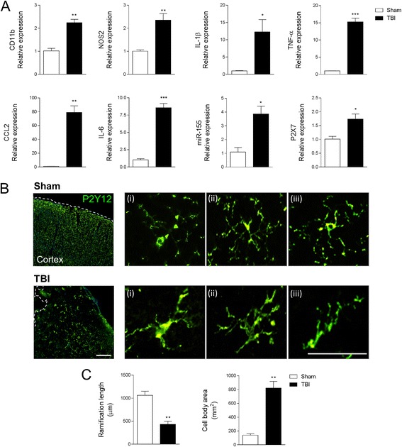
Adult male C57BL/6 mice were subjected to controlled cortical impact TBI for 24 h, and enriched MP were isolated in the blood, while neuroinflammation was assessed in the TBI cortex. MP were characterized by flow cytometry, and MP content was assayed using gene and protein markers for pro-inflammatory mediators. Enriched MP co-cultured with BV2 or primary microglial cells were used for immune propagation assays. Enriched MP from BV2 microglia or CD11b-positive microglia from the TBI brain were stereotactically injected into the cortex of uninjured mice to evaluate MP-related seeding of neuroinflammation in vivo.

RESULTS

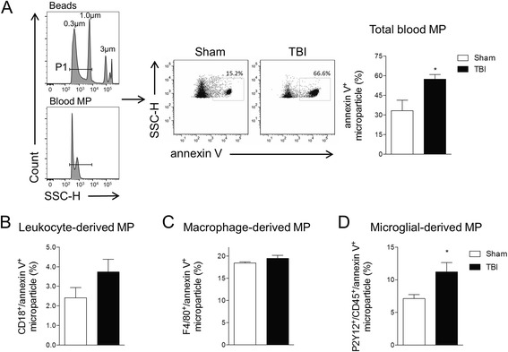
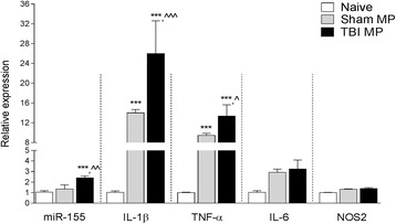
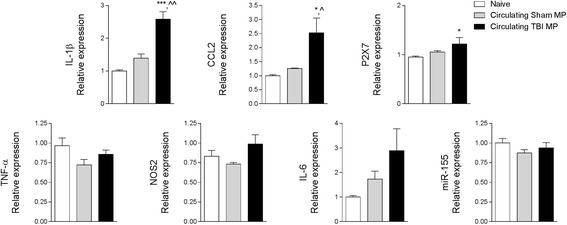
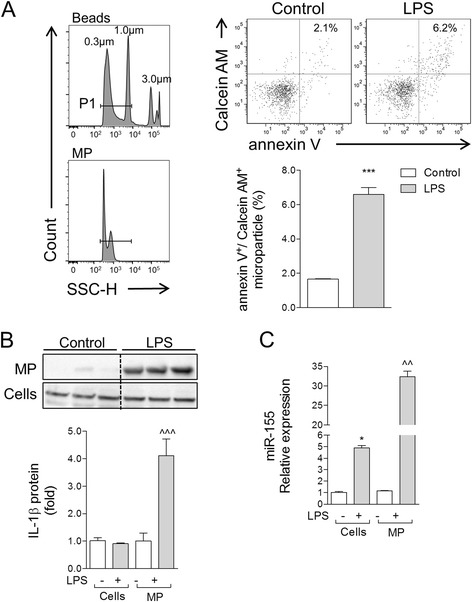
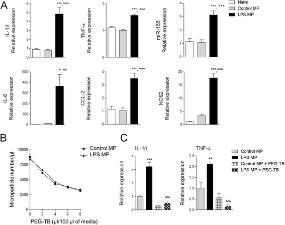
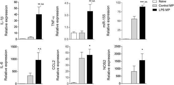
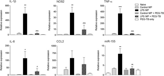
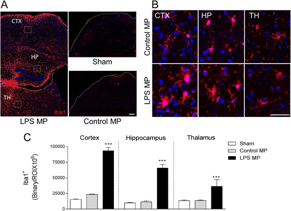
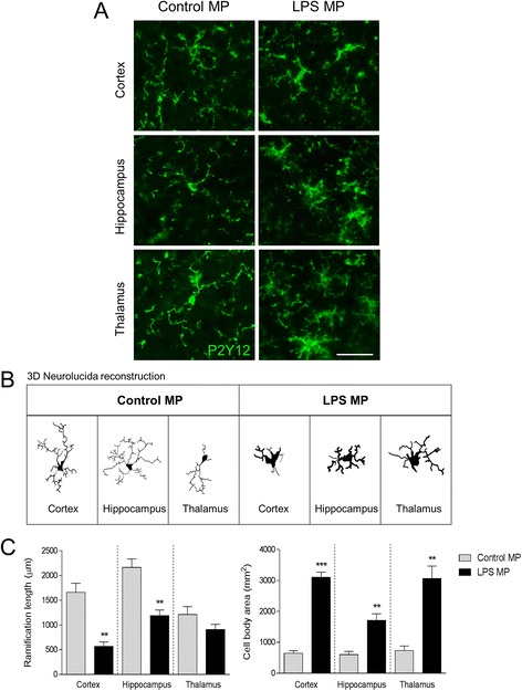
As the neuroinflammatory response is developing in the brain after TBI, microglial-derived MP are released into the circulation. Circulating enriched MP from the TBI animals can activate microglia in vitro. Lipopolysaccharide stimulation increases MP release from microglia in vitro and enhances their content of pro-inflammatory mediators, interleukin-1β and microRNA-155. Enriched MP from activated microglia in vitro or CD11b-isolated microglia/macrophage from the TBI brain ex vivo are sufficient to initiate neuroinflammation following their injection into the cortex of naïve (uninjured) animals.

CONCLUSIONS

These data provide further insights into the mechanisms underlying the development and dissemination of neuroinflammation after TBI. MP loaded with pro-inflammatory molecules initially released by microglia following trauma can activate additional microglia that may contribute to progressive neuroinflammatory response in the injured brain, as well as stimulate systemic immune responses. Due to their ability to independently initiate inflammatory responses, MP derived from activated microglia may provide a potential therapeutic target for other neurological disorders in which neuroinflammation may be a contributing factor.

摘要

背景

创伤性脑损伤(TBI)后早期即会引发局部和全身炎症反应,且可能在导致神经元丢失和神经功能缺损的继发性损伤过程中起关键作用。然而,神经炎症快速扩展及其长期进展的机制尚未阐明。在此,我们研究细胞外囊泡家族成员微颗粒(MP)在脑免疫细胞间促炎分子交换以及它们向体循环转移过程中的作用,将其作为脑外伤后炎症传播的关键途径。

方法

成年雄性C57BL/6小鼠接受控制性皮质撞击TBI 24小时,从血液中分离富集MP,同时评估TBI皮质中的神经炎症。通过流式细胞术对MP进行表征,并使用促炎介质的基因和蛋白质标志物检测MP含量。将富集的MP与BV2或原代小胶质细胞共培养用于免疫传播分析。将来自BV2小胶质细胞或TBI脑的CD11b阳性小胶质细胞的富集MP立体定向注射到未受伤小鼠的皮质中,以评估体内MP相关的神经炎症播种。

结果

TBI后随着脑内神经炎症反应的发展,小胶质细胞衍生的MP释放到循环中。来自TBI动物的循环富集MP可在体外激活小胶质细胞。脂多糖刺激可增加体外小胶质细胞的MP释放,并提高其促炎介质白细胞介素-1β和微小RNA-155的含量。体外活化小胶质细胞或TBI脑离体分离的CD11b小胶质细胞/巨噬细胞的富集MP注射到未受伤(天真的)动物皮质后足以引发神经炎症。

结论

这些数据为TBI后神经炎症发生和传播的潜在机制提供了进一步的见解。创伤后最初由小胶质细胞释放的载有促炎分子的MP可激活其他小胶质细胞,这可能导致受伤脑内进行性神经炎症反应,并刺激全身免疫反应。由于其独立引发炎症反应的能力,活化小胶质细胞衍生的MP可能为神经炎症可能是一个促成因素的其他神经系统疾病提供潜在的治疗靶点。

https://cdn.ncbi.nlm.nih.gov/pmc/blobs/e74c/5351060/c7a3bb816c86/12974_2017_819_Fig10_HTML.jpg
https://cdn.ncbi.nlm.nih.gov/pmc/blobs/e74c/5351060/c2c8f51e9197/12974_2017_819_Fig1_HTML.jpg
https://cdn.ncbi.nlm.nih.gov/pmc/blobs/e74c/5351060/86021293c787/12974_2017_819_Fig2_HTML.jpg
https://cdn.ncbi.nlm.nih.gov/pmc/blobs/e74c/5351060/bc8509c0cd0b/12974_2017_819_Fig3_HTML.jpg
https://cdn.ncbi.nlm.nih.gov/pmc/blobs/e74c/5351060/3bd8f8cd462c/12974_2017_819_Fig4_HTML.jpg
https://cdn.ncbi.nlm.nih.gov/pmc/blobs/e74c/5351060/7b52613d1936/12974_2017_819_Fig5_HTML.jpg
https://cdn.ncbi.nlm.nih.gov/pmc/blobs/e74c/5351060/17900a4f3170/12974_2017_819_Fig6_HTML.jpg
https://cdn.ncbi.nlm.nih.gov/pmc/blobs/e74c/5351060/29483eef3582/12974_2017_819_Fig7_HTML.jpg
https://cdn.ncbi.nlm.nih.gov/pmc/blobs/e74c/5351060/c2371ba4d7df/12974_2017_819_Fig8_HTML.jpg
https://cdn.ncbi.nlm.nih.gov/pmc/blobs/e74c/5351060/52c861ff8d8e/12974_2017_819_Fig9_HTML.jpg
https://cdn.ncbi.nlm.nih.gov/pmc/blobs/e74c/5351060/c7a3bb816c86/12974_2017_819_Fig10_HTML.jpg

相似文献

1
Microglial-derived microparticles mediate neuroinflammation after traumatic brain injury.小胶质细胞衍生的微粒在创伤性脑损伤后介导神经炎症。
J Neuroinflammation. 2017 Mar 15;14(1):47. doi: 10.1186/s12974-017-0819-4.
2
Omega-3 polyunsaturated fatty acid supplementation attenuates microglial-induced inflammation by inhibiting the HMGB1/TLR4/NF-κB pathway following experimental traumatic brain injury.实验性创伤性脑损伤后,补充ω-3多不饱和脂肪酸通过抑制HMGB1/TLR4/NF-κB信号通路减轻小胶质细胞诱导的炎症反应。
J Neuroinflammation. 2017 Jul 24;14(1):143. doi: 10.1186/s12974-017-0917-3.
3
Omega-3 polyunsaturated fatty acid attenuates the inflammatory response by modulating microglia polarization through SIRT1-mediated deacetylation of the HMGB1/NF-κB pathway following experimental traumatic brain injury.ω-3 多不饱和脂肪酸通过 SIRT1 介导的 HMGB1/NF-κB 通路的去乙酰化作用调节小胶质细胞极化,减轻实验性创伤性脑损伤后的炎症反应。
J Neuroinflammation. 2018 Apr 20;15(1):116. doi: 10.1186/s12974-018-1151-3.
4
Microglial Depletion with CSF1R Inhibitor During Chronic Phase of Experimental Traumatic Brain Injury Reduces Neurodegeneration and Neurological Deficits.实验性创伤性脑损伤慢性期使用 CSF1R 抑制剂耗竭小胶质细胞可减少神经退行性变和神经功能缺损。
J Neurosci. 2020 Apr 1;40(14):2960-2974. doi: 10.1523/JNEUROSCI.2402-19.2020. Epub 2020 Feb 24.
5
Increased miR-124-3p in microglial exosomes following traumatic brain injury inhibits neuronal inflammation and contributes to neurite outgrowth their transfer into neurons.创伤性脑损伤后小胶质细胞外泌体中 miR-124-3p 的增加抑制神经元炎症,促进轴突生长及其向神经元的转移。
FASEB J. 2018 Jan;32(1):512-528. doi: 10.1096/fj.201700673R. Epub 2017 Sep 21.
6
ACT001 attenuates microglia-mediated neuroinflammation after traumatic brain injury via inhibiting AKT/NFκB/NLRP3 pathway.ACT001 通过抑制 AKT/NFκB/NLRP3 通路来减轻创伤性脑损伤后的小胶质细胞介导的神经炎症。
Cell Commun Signal. 2022 Apr 23;20(1):56. doi: 10.1186/s12964-022-00862-y.
7
Traumatic brain injury-induced neuronal damage in the somatosensory cortex causes formation of rod-shaped microglia that promote astrogliosis and persistent neuroinflammation.创伤性脑损伤引起的感觉皮层神经元损伤导致杆状小胶质细胞的形成,促进星形胶质细胞增生和持续的神经炎症。
Glia. 2018 Dec;66(12):2719-2736. doi: 10.1002/glia.23523. Epub 2018 Oct 30.
8
Suppression of miR-193a alleviates neuroinflammation and improves neurological function recovery after traumatic brain injury (TBI) in mice.miR-193a 的抑制可减轻创伤性脑损伤 (TBI) 后小鼠的神经炎症并改善神经功能恢复。
Biochem Biophys Res Commun. 2020 Mar 5;523(2):527-534. doi: 10.1016/j.bbrc.2019.11.095. Epub 2020 Jan 8.
9
Progranulin protects against exaggerated axonal injury and astrogliosis following traumatic brain injury.颗粒蛋白前体可预防创伤性脑损伤后过度的轴突损伤和星形胶质细胞增生。
Glia. 2017 Feb;65(2):278-292. doi: 10.1002/glia.23091. Epub 2016 Oct 25.
10
Disruption of Midkine gene reduces traumatic brain injury through the modulation of neuroinflammation.Midkine 基因缺失通过调节神经炎症减轻创伤性脑损伤。
J Neuroinflammation. 2020 Jan 29;17(1):40. doi: 10.1186/s12974-020-1709-8.

引用本文的文献

1
Chrysin-Loaded Extracellular Vesicles Attenuate LPS-Induced Neuroinflammation in BV2 Microglial Cells In Vitro: A Novel Neuroprotective Strategy.载白杨素的细胞外囊泡体外减轻脂多糖诱导的BV2小胶质细胞神经炎症:一种新型神经保护策略。
Molecules. 2025 Jul 25;30(15):3131. doi: 10.3390/molecules30153131.
2
Stem cell-derived extracellular vesicles: novel therapeutics for cerebral injury following cardiac arrest and potential mechanisms.干细胞衍生的细胞外囊泡:心脏骤停后脑损伤的新型疗法及潜在机制
Cell Biosci. 2025 Jul 26;15(1):110. doi: 10.1186/s13578-025-01451-5.
3
Extracellular vesicles as a biomarkers in traumatic brain injury: a systematic review of animal and clinical studies.

本文引用的文献

1
In vivo inhibition of miR-155 significantly alters post-stroke inflammatory response.体内抑制miR-155可显著改变中风后的炎症反应。
J Neuroinflammation. 2016 Nov 9;13(1):287. doi: 10.1186/s12974-016-0753-x.
2
Humanizing the Protease-Activated Receptor (PAR) Expression Profile in Mouse Platelets by Knocking PAR1 into the Par3 Locus Reveals PAR1 Expression Is Not Tolerated in Mouse Platelets.通过将PAR1敲入Par3基因座使小鼠血小板中的蛋白酶激活受体(PAR)表达谱人源化,揭示了PAR1在小鼠血小板中不能被耐受。
PLoS One. 2016 Oct 27;11(10):e0165565. doi: 10.1371/journal.pone.0165565. eCollection 2016.
3
Microparticle formation by platelets exposed to high gas pressures - An oxidative stress response.
细胞外囊泡作为创伤性脑损伤的生物标志物:对动物和临床研究的系统评价
Crit Care. 2025 Jul 24;29(1):324. doi: 10.1186/s13054-025-05477-6.
4
Mitochondria: the hidden engines of traumatic brain injury-driven neurodegeneration.线粒体:创伤性脑损伤所致神经退行性变的隐匿引擎
Front Cell Neurosci. 2025 May 9;19:1570596. doi: 10.3389/fncel.2025.1570596. eCollection 2025.
5
Gasdermin-D Genetic Knockout Reduces Inflammasome-Induced Disruption of the Gut-Brain Axis After Traumatic Brain Injury.Gasdermin-D基因敲除可减轻创伤性脑损伤后炎性小体诱导的肠-脑轴破坏。
Int J Mol Sci. 2025 Apr 9;26(8):3512. doi: 10.3390/ijms26083512.
6
A Chloroform Fraction Derived from Root Ethanol Extract Attenuates Lipopolysaccharide-Induced Inflammatory Responses and Cognitive Dysfunction in BV-2 Microglia Cells and C57BL/6J Mouse Model.从根部乙醇提取物中获得的氯仿馏分可减轻脂多糖诱导的BV-2小胶质细胞和C57BL/6J小鼠模型中的炎症反应和认知功能障碍。
Int J Mol Sci. 2025 Mar 28;26(7):3126. doi: 10.3390/ijms26073126.
7
New role for an old acquaintance: miR-1246 as a new inflammatory and prognostic marker in polytrauma patients.老相识的新角色:miR-1246作为多发伤患者新的炎症和预后标志物
PeerJ. 2025 Apr 7;13:e19185. doi: 10.7717/peerj.19185. eCollection 2025.
8
Extracellular particles: emerging insights into central nervous system diseases.细胞外颗粒:对中枢神经系统疾病的新见解
J Nanobiotechnology. 2025 Apr 1;23(1):263. doi: 10.1186/s12951-025-03354-6.
9
Global status and trends of exosomes in neurodegenerative diseases from 2014 to 2023: a bibliometric and visual analysis.2014年至2023年神经退行性疾病中外泌体的全球现状与趋势:文献计量与可视化分析
Front Aging Neurosci. 2025 Mar 11;17:1496252. doi: 10.3389/fnagi.2025.1496252. eCollection 2025.
10
Enhanced exosome secretion regulated by microglial P2X7R in the medullary dorsal horn contributes to pulpitis-induced pain.髓质背角中由小胶质细胞P2X7R调节的外泌体分泌增强促成了牙髓炎诱导的疼痛。
Cell Biosci. 2025 Feb 22;15(1):28. doi: 10.1186/s13578-025-01363-4.
血小板在高压气体下形成微颗粒 - 一种氧化应激反应。
Free Radic Biol Med. 2016 Dec;101:154-162. doi: 10.1016/j.freeradbiomed.2016.10.010. Epub 2016 Oct 15.
4
Adipocyte-derived microvesicles from obese mice induce M1 macrophage phenotype through secreted miR-155.来自肥胖小鼠的脂肪细胞衍生微泡通过分泌的miR-155诱导M1巨噬细胞表型。
J Mol Cell Biol. 2016 Dec;8(6):505-517. doi: 10.1093/jmcb/mjw040. Epub 2016 Sep 25.
5
Traumatic brain injury increases levels of miR-21 in extracellular vesicles: implications for neuroinflammation.创伤性脑损伤会增加细胞外囊泡中miR-21的水平:对神经炎症的影响。
FEBS Open Bio. 2016 Jun 14;6(8):835-46. doi: 10.1002/2211-5463.12092. eCollection 2016 Aug.
6
NOX2 drives M1-like microglial/macrophage activation and neurodegeneration following experimental traumatic brain injury.在实验性创伤性脑损伤后,NOX2驱动M1样小胶质细胞/巨噬细胞活化和神经退行性变。
Brain Behav Immun. 2016 Nov;58:291-309. doi: 10.1016/j.bbi.2016.07.158. Epub 2016 Jul 28.
7
Extracellular vesicles and intercellular communication within the nervous system.细胞外囊泡与神经系统内的细胞间通讯。
J Clin Invest. 2016 Apr 1;126(4):1198-207. doi: 10.1172/JCI81134.
8
Endothelial microparticles carrying hedgehog-interacting protein induce continuous endothelial damage in the pathogenesis of acute graft-versus-host disease.携带刺猬因子相互作用蛋白的内皮微粒在急性移植物抗宿主病发病机制中诱导持续性内皮损伤。
Am J Physiol Cell Physiol. 2016 May 15;310(10):C821-35. doi: 10.1152/ajpcell.00372.2015. Epub 2016 Mar 23.
9
Mechanical Injury Induces Brain Endothelial-Derived Microvesicle Release: Implications for Cerebral Vascular Injury during Traumatic Brain Injury.机械损伤诱导脑内皮源性微泡释放:对创伤性脑损伤期间脑血管损伤的影响。
Front Cell Neurosci. 2016 Feb 29;10:43. doi: 10.3389/fncel.2016.00043. eCollection 2016.
10
Communication by Extracellular Vesicles: Where We Are and Where We Need to Go.细胞外囊泡通讯:我们的现状与未来展望。
Cell. 2016 Mar 10;164(6):1226-1232. doi: 10.1016/j.cell.2016.01.043.