• 文献检索
  • 文档翻译
  • 深度研究
  • 学术资讯
  • Suppr Zotero 插件Zotero 插件
  • 邀请有礼
  • 套餐&价格
  • 历史记录
应用&插件
Suppr Zotero 插件Zotero 插件浏览器插件Mac 客户端Windows 客户端微信小程序
定价
高级版会员购买积分包购买API积分包
服务
文献检索文档翻译深度研究API 文档MCP 服务
关于我们
关于 Suppr公司介绍联系我们用户协议隐私条款
关注我们

Suppr 超能文献

核心技术专利:CN118964589B侵权必究
粤ICP备2023148730 号-1Suppr @ 2026

文献检索

告别复杂PubMed语法,用中文像聊天一样搜索,搜遍4000万医学文献。AI智能推荐,让科研检索更轻松。

立即免费搜索

文件翻译

保留排版,准确专业,支持PDF/Word/PPT等文件格式,支持 12+语言互译。

免费翻译文档

深度研究

AI帮你快速写综述,25分钟生成高质量综述,智能提取关键信息,辅助科研写作。

立即免费体验

基于基线 CT 筛查结节的恶性风险评估:PanCan 模型、Lung-RADS 和 NCCN 指南的比较。

Malignancy risk estimation of screen-detected nodules at baseline CT: comparison of the PanCan model, Lung-RADS and NCCN guidelines.

机构信息

Department of Radiology and Nuclear Medicine, Radboud University Nijmegen Medical Center, Nijmegen, The Netherlands.

Department of Pulmonology Gentofte Hospital, University of Copenhagen, Hellerup, Denmark.

出版信息

Eur Radiol. 2017 Oct;27(10):4019-4029. doi: 10.1007/s00330-017-4767-2. Epub 2017 Mar 14.

DOI:10.1007/s00330-017-4767-2
PMID:28293773
原文链接:https://pmc.ncbi.nlm.nih.gov/articles/PMC5579178/
Abstract

OBJECTIVES

To compare the PanCan model, Lung-RADS and the 1.2016 National Comprehensive Cancer Network (NCCN) guidelines for discriminating malignant from benign pulmonary nodules on baseline screening CT scans and the impact diameter measurement methods have on performances.

METHODS

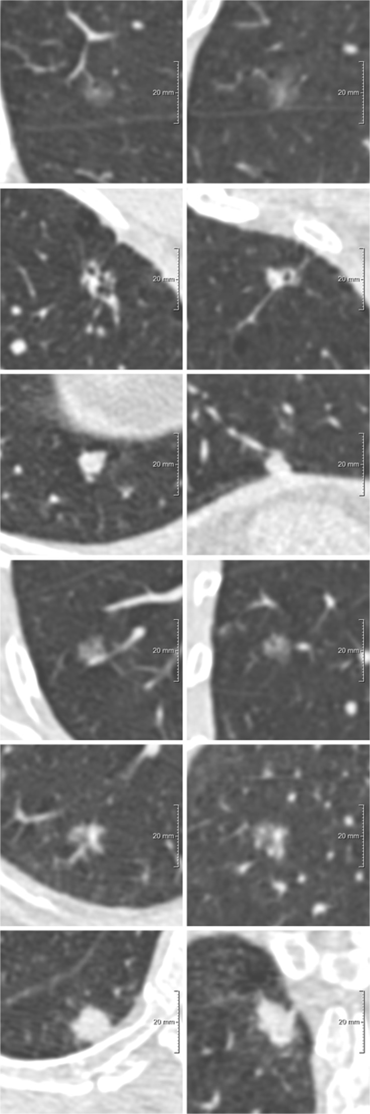
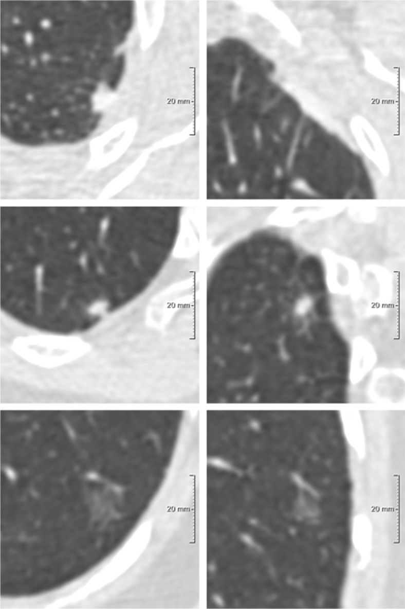
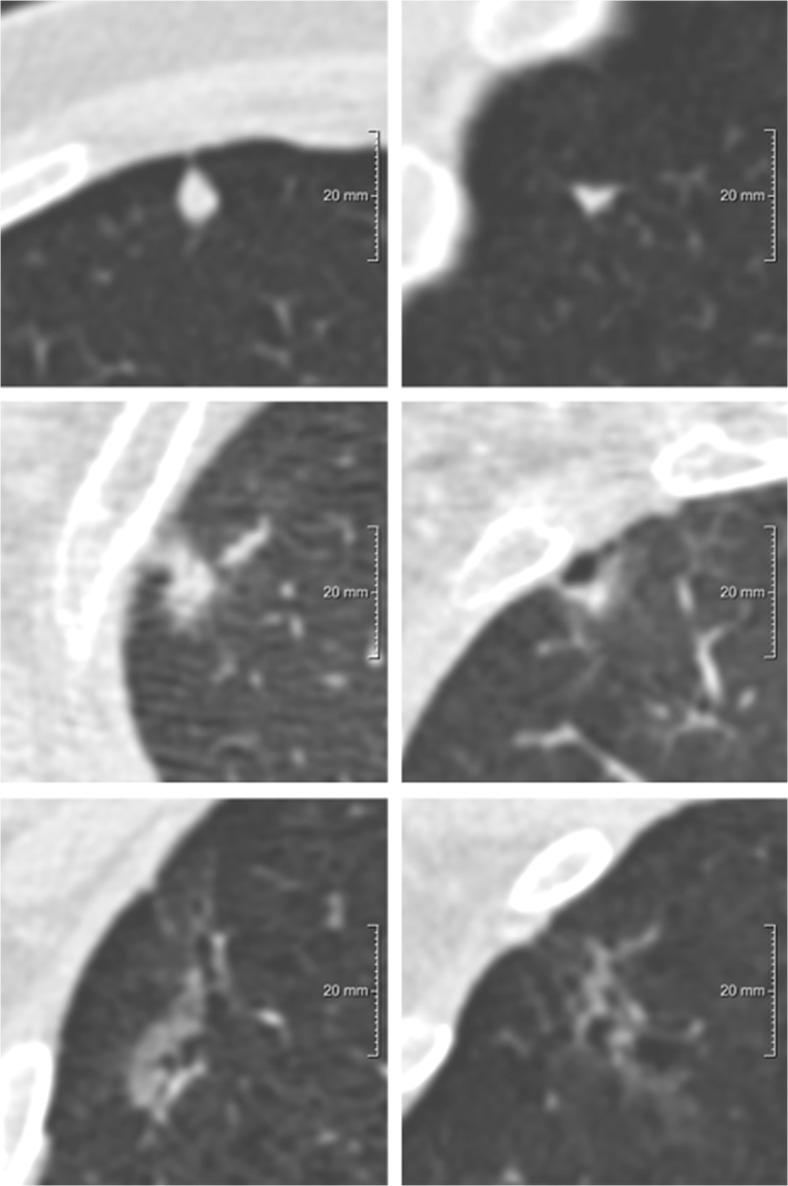
From the Danish Lung Cancer Screening Trial database, 64 CTs with malignant nodules and 549 baseline CTs with benign nodules were included. Performance of the systems was evaluated applying the system's original diameter definitions: D (PanCan), D (NCCN), both obtained from axial sections, and D (Lung-RADS). Subsequently all diameter definitions were applied uniformly to all systems. Areas under the ROC curves (AUC) were used to evaluate risk discrimination.

RESULTS

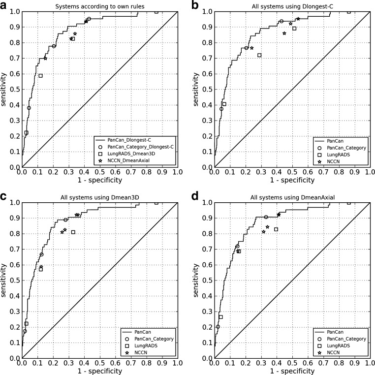
PanCan performed superiorly to Lung-RADS and NCCN (AUC 0.874 vs. 0.813, p = 0.003; 0.874 vs. 0.836, p = 0.010), using the original diameter specifications. When uniformly applying D, D and D, PanCan remained superior to Lung-RADS (p < 0.001 - p = 0.001) and NCCN (p < 0.001 - p = 0.016). Diameter definition significantly influenced NCCN's performance with D being the worst (D vs. D, p = 0.005; D vs. D, p = 0.016).

CONCLUSIONS

Without follow-up information, the PanCan model performs significantly superiorly to Lung-RADS and the 1.2016 NCCN guidelines for discriminating benign from malignant nodules. The NCCN guidelines are most sensitive to nodule size definition.

KEY POINTS

• PanCan model outperforms Lung-RADS and 1.2016 NCCN guidelines in identifying malignant pulmonary nodules. • Nodule size definition had no significant impact on Lung-RADS and PanCan model. • 1.2016 NCCN guidelines were significantly superior when using mean diameter to longest diameter. • Longest diameter achieved lowest performance for all models. • Mean diameter performed equivalently when derived from axial sections and from volumetry.

摘要

目的

比较 PanCan 模型、Lung-RADS 和 2016 年 1 月国家综合癌症网络(NCCN)指南在基线筛查 CT 扫描中鉴别良恶性肺结节的性能,以及直径测量方法对性能的影响。

方法

从丹麦肺癌筛查试验数据库中纳入 64 例恶性结节 CT 和 549 例基线良性结节 CT。应用系统的原始直径定义评估系统的性能:D(PanCan)、D(NCCN),均从轴向切片获得,以及 D(Lung-RADS)。随后,所有直径定义均应用于所有系统。受试者工作特征曲线下面积(AUC)用于评估风险判别。

结果

PanCan 模型的性能优于 Lung-RADS 和 NCCN(AUC 0.874 比 0.813,p=0.003;0.874 比 0.836,p=0.010),使用原始直径规格。当均匀应用 D、D 和 D 时,PanCan 仍优于 Lung-RADS(p<0.001-p=0.001)和 NCCN(p<0.001-p=0.016)。直径定义显著影响 NCCN 的性能,D 最差(D 比 D,p=0.005;D 比 D,p=0.016)。

结论

无随访信息时,PanCan 模型在鉴别良恶性结节方面的性能明显优于 Lung-RADS 和 2016 年 1 月 NCCN 指南。NCCN 指南对结节大小定义最敏感。

要点

• PanCan 模型在识别恶性肺结节方面优于 Lung-RADS 和 2016 年 1 月 NCCN 指南。• 结节大小定义对 Lung-RADS 和 PanCan 模型没有显著影响。• 使用平均直径与最长直径时,2016 年 1 月 NCCN 指南的性能明显优于其他指南。• 所有模型的最长直径性能最低。• 当源自轴向切片和体层时,平均直径的性能相当。

https://cdn.ncbi.nlm.nih.gov/pmc/blobs/4681/5579178/5f43a1f058d7/330_2017_4767_Fig4_HTML.jpg
https://cdn.ncbi.nlm.nih.gov/pmc/blobs/4681/5579178/073e2ea0deb9/330_2017_4767_Fig1_HTML.jpg
https://cdn.ncbi.nlm.nih.gov/pmc/blobs/4681/5579178/90e02d8d4368/330_2017_4767_Fig2_HTML.jpg
https://cdn.ncbi.nlm.nih.gov/pmc/blobs/4681/5579178/5f7ff44471ea/330_2017_4767_Fig3_HTML.jpg
https://cdn.ncbi.nlm.nih.gov/pmc/blobs/4681/5579178/5f43a1f058d7/330_2017_4767_Fig4_HTML.jpg

相似文献

1
Malignancy risk estimation of screen-detected nodules at baseline CT: comparison of the PanCan model, Lung-RADS and NCCN guidelines.基于基线 CT 筛查结节的恶性风险评估:PanCan 模型、Lung-RADS 和 NCCN 指南的比较。
Eur Radiol. 2017 Oct;27(10):4019-4029. doi: 10.1007/s00330-017-4767-2. Epub 2017 Mar 14.
2
Evaluation of Prediction Models for Identifying Malignancy in Pulmonary Nodules Detected via Low-Dose Computed Tomography.基于低剂量 CT 检测肺结节的良恶性鉴别预测模型评估。
JAMA Netw Open. 2020 Feb 5;3(2):e1921221. doi: 10.1001/jamanetworkopen.2019.21221.
3
A Comparison of the PanCan Model and Lung-RADS to Assess Cancer Probability Among People With Screening-Detected, Solid Lung Nodules.一种比较 PanCan 模型和 Lung-RADS 在评估有筛查发现的实性肺结节人群中的癌症可能性的方法。
Chest. 2021 Mar;159(3):1273-1282. doi: 10.1016/j.chest.2020.10.040. Epub 2020 Oct 23.
4
Predicting Malignancy Risk of Screen-Detected Lung Nodules-Mean Diameter or Volume.基于直径或体积预测筛查性肺结节的恶性风险
J Thorac Oncol. 2019 Feb;14(2):203-211. doi: 10.1016/j.jtho.2018.10.006. Epub 2018 Oct 25.
5
Deep Learning for Malignancy Risk Estimation of Pulmonary Nodules Detected at Low-Dose Screening CT.基于低剂量 CT 扫描检测到的肺部结节的恶性肿瘤风险估计的深度学习。
Radiology. 2021 Aug;300(2):438-447. doi: 10.1148/radiol.2021204433. Epub 2021 May 18.
6
Malignancy risk estimation of pulmonary nodules in screening CTs: Comparison between a computer model and human observers.筛查CT中肺结节的恶性风险评估:计算机模型与人类观察者的比较。
PLoS One. 2017 Nov 9;12(11):e0185032. doi: 10.1371/journal.pone.0185032. eCollection 2017.
7
Probability of cancer in pulmonary nodules detected on first screening CT.首次筛查 CT 检测到的肺结节的癌症概率。
N Engl J Med. 2013 Sep 5;369(10):910-9. doi: 10.1056/NEJMoa1214726.
8
Screen-detected subsolid pulmonary nodules: long-term follow-up and application of the PanCan lung cancer risk prediction model.筛查发现的亚实性肺结节:长期随访及泛癌种肺癌风险预测模型的应用
Br J Radiol. 2016;89(1060):20160016. doi: 10.1259/bjr.20160016. Epub 2016 Feb 16.
9
Malignancy estimation of Lung-RADS criteria for subsolid nodules on CT: accuracy of low and high risk spectrum when using NLST nodules.CT 上亚实性结节 Lung-RADS 标准的恶性估计:使用 NLST 结节时低风险和高风险谱的准确性。
Eur Radiol. 2017 Nov;27(11):4672-4679. doi: 10.1007/s00330-017-4842-8. Epub 2017 Apr 24.
10
Predictive Accuracy of the PanCan Lung Cancer Risk Prediction Model -External Validation based on CT from the Danish Lung Cancer Screening Trial.泛癌种肺癌风险预测模型的预测准确性——基于丹麦肺癌筛查试验CT的外部验证
Eur Radiol. 2015 Oct;25(10):3093-9. doi: 10.1007/s00330-015-3689-0. Epub 2015 Mar 13.

引用本文的文献

1
Diagnostic and therapeutic strategies for solitary pulmonary nodules mimicking malignancy: Insights from two cases of pulmonary tuberculosis.疑似恶性的孤立性肺结节的诊断和治疗策略:两例肺结核病例的启示
IDCases. 2025 Jun 23;41:e02302. doi: 10.1016/j.idcr.2025.e02302. eCollection 2025.
2
Lung Cancer Risk Prediction in Patients with Persistent Pulmonary Nodules Using the Brock Model and Sybil Model.使用布罗克模型和西比尔模型预测持续性肺结节患者的肺癌风险
Cancers (Basel). 2025 Apr 29;17(9):1499. doi: 10.3390/cancers17091499.
3
Clinical effect of Almonertinib in treating epidermal growth factor receptor mutation-positive residual ground-glass opacities after stage I lung cancer resection.

本文引用的文献

1
Development of Guidelines for the Management of Pulmonary Nodules: Toward Better Implementation.肺结节管理指南的制定:迈向更好的实施
Chest. 2015 Dec;148(6):1365-1367. doi: 10.1378/chest.15-1906.
2
Results of the Randomized Danish Lung Cancer Screening Trial with Focus on High-Risk Profiling.随机丹麦肺癌筛查试验的结果,重点关注高危人群特征。
Am J Respir Crit Care Med. 2016 Mar 1;193(5):542-51. doi: 10.1164/rccm.201505-1040OC.
3
Persistent pulmonary subsolid nodules with solid portions of 5 mm or smaller: Their natural course and predictors of interval growth.
阿美替尼治疗Ⅰ期肺癌切除术后表皮生长因子受体突变阳性残留磨玻璃影的临床疗效
Am J Transl Res. 2024 Dec 15;16(12):7553-7562. doi: 10.62347/TOYK7025. eCollection 2024.
4
New developments in the imaging of lung cancer.肺癌成像的新进展。
Breathe (Sheff). 2024 Mar;20(1):230176. doi: 10.1183/20734735.0176-2023. Epub 2024 Apr 9.
5
Computed tomography-guided biopsy for sub-centimetre pulmonary nodules: a meta-analysis.计算机断层扫描引导下的亚厘米级肺结节活检:一项荟萃分析。
Kardiochir Torakochirurgia Pol. 2023 Sep;20(3):139-145. doi: 10.5114/kitp.2023.131947. Epub 2023 Oct 30.
6
Clinical Adjuncts to Lung Cancer Screening: A Narrative Review.肺癌筛查的临床辅助手段:一篇叙述性综述。
Thorac Surg Clin. 2023 Nov;33(4):421-432. doi: 10.1016/j.thorsurg.2023.03.002. Epub 2023 May 12.
7
Developing an understanding of artificial intelligence lung nodule risk prediction using insights from the Brock model.利用 Brock 模型的见解来理解人工智能肺结节风险预测。
Eur Radiol. 2022 Aug;32(8):5330-5338. doi: 10.1007/s00330-022-08635-4. Epub 2022 Mar 3.
8
Ultrashort echo time MRI of the lung in children and adolescents: comparison with non-enhanced computed tomography and standard post-contrast T1w MRI sequences.儿童和青少年肺部超短回波时间 MRI:与非增强 CT 及标准对比增强 T1w MRI 序列的比较。
Eur Radiol. 2022 Mar;32(3):1833-1842. doi: 10.1007/s00330-021-08236-7. Epub 2021 Oct 20.
9
Contemporary issues in the implementation of lung cancer screening.肺癌筛查实施中的当代问题。
Eur Respir Rev. 2021 Jul 20;30(161). doi: 10.1183/16000617.0288-2020. Print 2021 Sep 30.
10
Artificial intelligence for detection and characterization of pulmonary nodules in lung cancer CT screening: ready for practice?人工智能在肺癌CT筛查中检测和表征肺结节:准备好应用于临床了吗?
Transl Lung Cancer Res. 2021 May;10(5):2378-2388. doi: 10.21037/tlcr-2020-lcs-06.
实性部分为5毫米或更小的持续性肺部亚实性结节:其自然病程及随访期生长的预测因素
Eur Radiol. 2016 Jun;26(6):1529-37. doi: 10.1007/s00330-015-4017-4. Epub 2015 Sep 18.
4
CT Screening for Lung Cancer: Nonsolid Nodules in Baseline and Annual Repeat Rounds.CT筛查肺癌:基线及年度复查中的非实性结节
Radiology. 2015 Nov;277(2):555-64. doi: 10.1148/radiol.2015142554. Epub 2015 Jun 23.
5
British Thoracic Society guidelines for the investigation and management of pulmonary nodules.英国胸科学会肺结节的调查与管理指南。
Thorax. 2015 Aug;70 Suppl 2:ii1-ii54. doi: 10.1136/thoraxjnl-2015-207168.
6
Critique of Al-Ameri et al. (2015) - Risk of malignancy in pulmonary nodules: A validation study of four prediction models.对阿勒梅里等人(2015年)的评论——肺结节的恶性风险:四种预测模型的验证研究
Lung Cancer. 2015 Oct;90(1):118-9. doi: 10.1016/j.lungcan.2015.05.015. Epub 2015 Jun 4.
7
Observer Variability for Classification of Pulmonary Nodules on Low-Dose CT Images and Its Effect on Nodule Management.低剂量 CT 图像上肺结节分类的观察者变异性及其对结节管理的影响。
Radiology. 2015 Dec;277(3):863-71. doi: 10.1148/radiol.2015142700. Epub 2015 May 22.
8
Predictive Accuracy of the PanCan Lung Cancer Risk Prediction Model -External Validation based on CT from the Danish Lung Cancer Screening Trial.泛癌种肺癌风险预测模型的预测准确性——基于丹麦肺癌筛查试验CT的外部验证
Eur Radiol. 2015 Oct;25(10):3093-9. doi: 10.1007/s00330-015-3689-0. Epub 2015 Mar 13.
9
Lung cancer probability in patients with CT-detected pulmonary nodules: a prespecified analysis of data from the NELSON trial of low-dose CT screening.CT 检测到肺结节患者的肺癌概率:来自低剂量 CT 筛查 NELSON 试验数据的预设分析。
Lancet Oncol. 2014 Nov;15(12):1332-41. doi: 10.1016/S1470-2045(14)70389-4. Epub 2014 Oct 1.
10
Resource utilization and costs during the initial years of lung cancer screening with computed tomography in Canada.加拿大肺癌 CT 筛查初始阶段的资源利用和成本。
J Thorac Oncol. 2014 Oct;9(10):1449-58. doi: 10.1097/JTO.0000000000000283.