Wan Bing, Wang Siqi, Tu Mengqi, Wu Bo, Han Ping, Xu Haibo
Department of Radiology, Union Hospital of Tongji Medical College, Huazhong University of Science and Technology Department of Radiology, Zhongnan Hospital of Wuhan University, Wuhan, China.
Medicine (Baltimore). 2017 Mar;96(11):e6333. doi: 10.1097/MD.0000000000006333.
The purpose of this meta-analysis was to evaluate the diagnostic accuracy of perfusion magnetic resonance imaging (MRI) as a method for differentiating glioma recurrence from pseudoprogression.
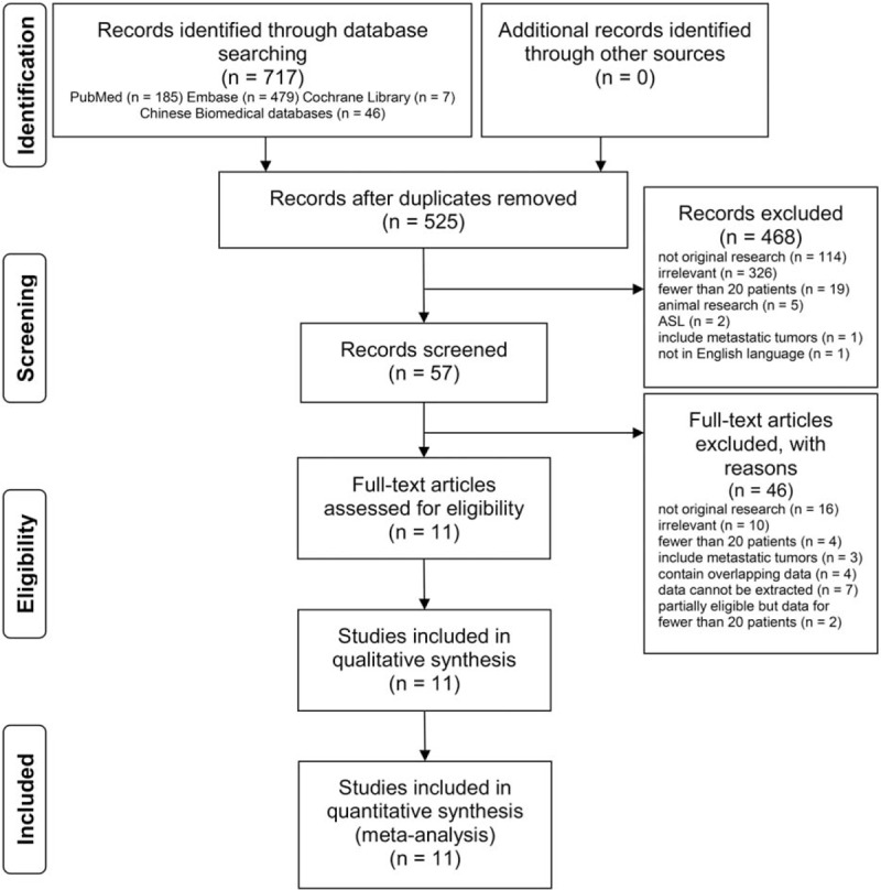
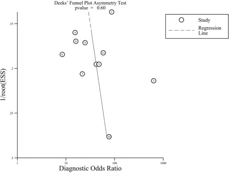
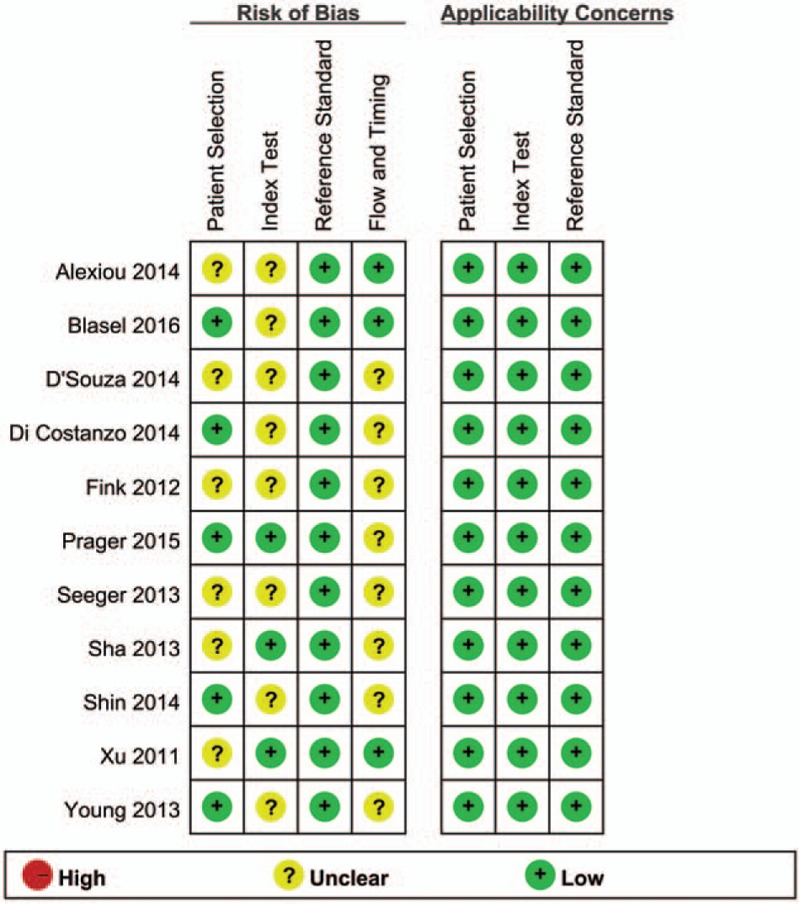
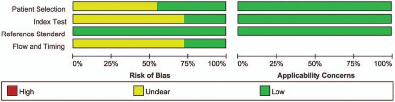
The PubMed, Embase, Cochrane Library, and Chinese Biomedical databases were searched comprehensively for relevant studies up to August 3, 2016 according to specific inclusion and exclusion criteria. The quality of the included studies was assessed according to the quality assessment of diagnostic accuracy studies (QUADAS-2). After performing heterogeneity and threshold effect tests, pooled sensitivity, specificity, positive likelihood ratio, negative likelihood ratio, and diagnostic odds ratio were calculated. Publication bias was evaluated visually by a funnel plot and quantitatively using Deek funnel plot asymmetry test. The area under the summary receiver operating characteristic curve was calculated to demonstrate the diagnostic performance of perfusion MRI.
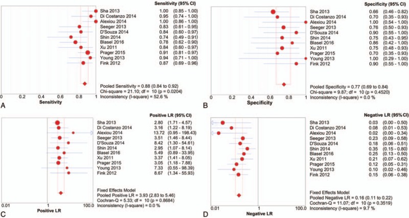
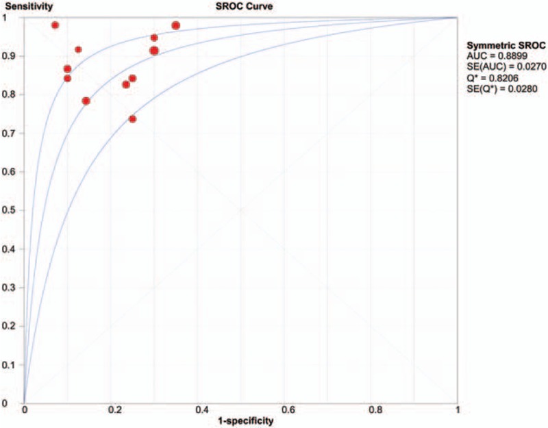
Eleven studies covering 416 patients and 418 lesions were included in this meta-analysis. The pooled sensitivity, specificity, positive likelihood ratio, negative likelihood ratio, and diagnostic odds ratio were 0.88 (95% confidence interval [CI] 0.84-0.92), 0.77 (95% CI 0.69-0.84), 3.93 (95% CI 2.83-5.46), 0.16 (95% CI 0.11-0.22), and 27.17 (95% CI 14.96-49.35), respectively. The area under the summary receiver operating characteristic curve was 0.8899. There was no notable publication bias. Sensitivity analysis showed that the meta-analysis results were stable and credible.
While perfusion MRI is not the ideal diagnostic method for differentiating glioma recurrence from pseudoprogression, it could improve diagnostic accuracy. Therefore, further research on combining perfusion MRI with other imaging modalities is warranted.
本荟萃分析的目的是评估灌注磁共振成像(MRI)作为鉴别胶质瘤复发与假性进展方法的诊断准确性。
根据特定的纳入和排除标准,全面检索了截至2016年8月3日的PubMed、Embase、Cochrane图书馆和中国生物医学数据库中的相关研究。根据诊断准确性研究的质量评估(QUADAS-2)对纳入研究的质量进行评估。在进行异质性和阈值效应检验后,计算合并敏感性、特异性、阳性似然比、阴性似然比和诊断比值比。通过漏斗图直观评估发表偏倚,并使用Deek漏斗图不对称检验进行定量评估。计算汇总受试者工作特征曲线下面积以证明灌注MRI的诊断性能。
本荟萃分析纳入了11项研究,共416例患者和418个病灶。合并敏感性、特异性、阳性似然比、阴性似然比和诊断比值比分别为0.88(95%置信区间[CI]0.84-0.92)、0.77(95%CI0.69-0.84)、3.93(95%CI2.83-5.46)、0.16(95%CI0.11-0.22)和27.17(95%CI14.96-49.35)。汇总受试者工作特征曲线下面积为0.8899。没有明显的发表偏倚。敏感性分析表明荟萃分析结果稳定可靠。
虽然灌注MRI不是鉴别胶质瘤复发与假性进展的理想诊断方法,但它可以提高诊断准确性。因此,有必要进一步研究将灌注MRI与其他成像方式相结合。