• 文献检索
  • 文档翻译
  • 深度研究
  • 学术资讯
  • Suppr Zotero 插件Zotero 插件
  • 邀请有礼
  • 套餐&价格
  • 历史记录
应用&插件
Suppr Zotero 插件Zotero 插件浏览器插件Mac 客户端Windows 客户端微信小程序
定价
高级版会员购买积分包购买API积分包
服务
文献检索文档翻译深度研究API 文档MCP 服务
关于我们
关于 Suppr公司介绍联系我们用户协议隐私条款
关注我们

Suppr 超能文献

核心技术专利:CN118964589B侵权必究
粤ICP备2023148730 号-1Suppr @ 2026

文献检索

告别复杂PubMed语法,用中文像聊天一样搜索,搜遍4000万医学文献。AI智能推荐,让科研检索更轻松。

立即免费搜索

文件翻译

保留排版,准确专业,支持PDF/Word/PPT等文件格式,支持 12+语言互译。

免费翻译文档

深度研究

AI帮你快速写综述,25分钟生成高质量综述,智能提取关键信息,辅助科研写作。

立即免费体验

基于表观扩散系数测量的扩散磁共振成像鉴别胶质瘤复发与假性进展诊断性能的Meta分析

Meta-analysis of the diagnostic performance of diffusion magnetic resonance imaging with apparent diffusion coefficient measurements for differentiating glioma recurrence from pseudoprogression.

作者信息

Yu Yang, Ma Yue, Sun Mengyao, Jiang Wenyan, Yuan Tingting, Tong Dan

机构信息

Department of Radiology.

Department of Internal Oncology, The First Hospital of Jilin University, Changchun, Jilin, China.

出版信息

Medicine (Baltimore). 2020 Jun 5;99(23):e20270. doi: 10.1097/MD.0000000000020270.

DOI:10.1097/MD.0000000000020270
PMID:32501974
原文链接:https://pmc.ncbi.nlm.nih.gov/articles/PMC7306328/
Abstract

OBJECTIVE

The accurate differentiation of glioma recurrence from pseudoprogression (PSP) after therapy remains a considerable clinical challenge. Several studies have shown that diffusion magnetic resonance imaging (MRI) has potential value in distinguishing these 2 outcomes. The current meta-analysis examined the diagnostic accuracy of diffusion MRI with the apparent diffusion coefficient (ADC) in the differentiation of glioma recurrence from PSP.

METHOD

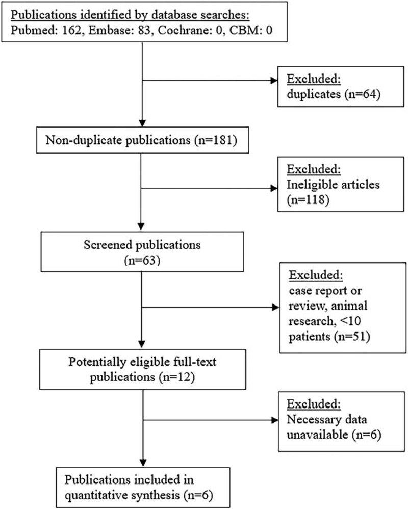
PubMed, Embase, Cochrane Library, and Chinese Biomedical databases were reviewed to identify studies that fulfilled our inclusion/exclusion criteria and were published on or before May 5, 2019. Threshold effects; heterogeneity; pooled sensitivity (SENS), specificity, positive likelihood ratio, and negative likelihood ratio; and diagnostic odds ratio were calculated. The overall diagnostic usefulness of diffusion MRI-derived ADC values was assessed by calculating the area under the curve (AUC) following summary receiver operating characteristic (SROC) analysis.

RESULTS

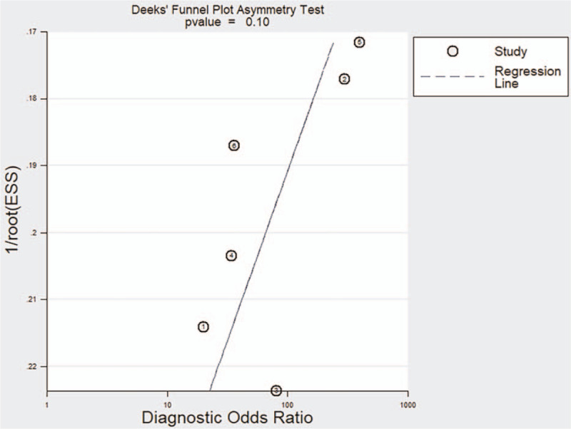
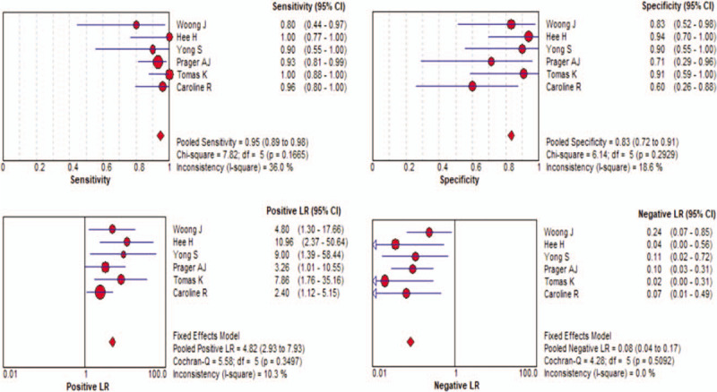
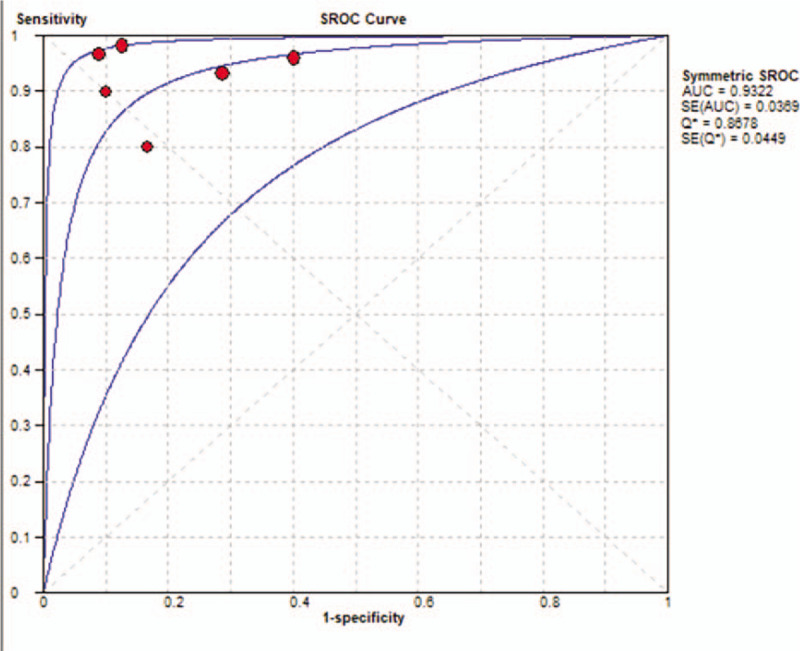
Six eligible studies examined a total of 214 patients. Calculation of pooled values indicated the SENS was 0.95 (95% confidence interval [CI] = 0.89-0.98), specificity was 0.83 (95% CI = 0.72-0.91), positive likelihood ratio was 4.82 (95% CI = 2.93-7.93), negative likelihood ratio was 0.08 (95% CI = 0.04-0.17), and diagnostic odds ratio was 59.63 (95% CI = 22.63-157.37). The SROC AUC was 0.9322. Publication bias was not significant, and SENS analysis indicated the results were relatively stable.

CONCLUSIONS

Our meta-analysis indicated that diffusion MRI with quantitative ADC is an effective approach for differentiation of glioma recurrence from PSP, and can be used as an auxiliary tool to diagnose glioma progression.

摘要

目的

准确区分治疗后胶质瘤复发与假性进展(PSP)仍是一项重大的临床挑战。多项研究表明,扩散磁共振成像(MRI)在区分这两种结果方面具有潜在价值。本荟萃分析探讨了基于表观扩散系数(ADC)的扩散MRI在鉴别胶质瘤复发与PSP中的诊断准确性。

方法

检索PubMed、Embase、Cochrane图书馆和中国生物医学数据库,以识别符合纳入/排除标准且于2019年5月5日或之前发表的研究。计算阈值效应、异质性、合并敏感度(SENS)、特异度、阳性似然比和阴性似然比以及诊断比值比。通过汇总接受者操作特征(SROC)分析计算曲线下面积(AUC),评估扩散MRI衍生的ADC值的总体诊断效用。

结果

六项符合条件的研究共纳入214例患者。合并值计算表明,SENS为0.95(95%置信区间[CI]=0.89-0.98),特异度为0.83(95%CI=0.72-0.91),阳性似然比为4.82(95%CI=2.93-7.93),阴性似然比为0.08(95%CI=0.04-0.17),诊断比值比为59.63(95%CI=22.63-157.37)。SROC AUC为0.9322。发表偏倚不显著,SENS分析表明结果相对稳定。

结论

我们的荟萃分析表明,基于定量ADC的扩散MRI是区分胶质瘤复发与PSP的有效方法,可作为诊断胶质瘤进展的辅助工具。

https://cdn.ncbi.nlm.nih.gov/pmc/blobs/90fd/7306328/0e7202b42814/medi-99-e20270-g007.jpg
https://cdn.ncbi.nlm.nih.gov/pmc/blobs/90fd/7306328/f01d20612ff4/medi-99-e20270-g002.jpg
https://cdn.ncbi.nlm.nih.gov/pmc/blobs/90fd/7306328/787d05d480d7/medi-99-e20270-g004.jpg
https://cdn.ncbi.nlm.nih.gov/pmc/blobs/90fd/7306328/eaec4fd10cd1/medi-99-e20270-g005.jpg
https://cdn.ncbi.nlm.nih.gov/pmc/blobs/90fd/7306328/9aa5000540e2/medi-99-e20270-g006.jpg
https://cdn.ncbi.nlm.nih.gov/pmc/blobs/90fd/7306328/0e7202b42814/medi-99-e20270-g007.jpg

相似文献

1
Meta-analysis of the diagnostic performance of diffusion magnetic resonance imaging with apparent diffusion coefficient measurements for differentiating glioma recurrence from pseudoprogression.基于表观扩散系数测量的扩散磁共振成像鉴别胶质瘤复发与假性进展诊断性能的Meta分析
Medicine (Baltimore). 2020 Jun 5;99(23):e20270. doi: 10.1097/MD.0000000000020270.
2
The diagnostic performance of perfusion MRI for differentiating glioma recurrence from pseudoprogression: A meta-analysis.灌注磁共振成像鉴别胶质瘤复发与假性进展的诊断效能:一项Meta分析。
Medicine (Baltimore). 2017 Mar;96(11):e6333. doi: 10.1097/MD.0000000000006333.
3
Diagnostic accuracy of dynamic contrast-enhanced magnetic resonance imaging for distinguishing pseudoprogression from glioma recurrence: a meta-analysis.动态对比增强磁共振成像鉴别假性进展与脑胶质瘤复发的诊断准确性:一项荟萃分析。
Chin Med J (Engl). 2021 Oct 14;134(21):2535-2543. doi: 10.1097/CM9.0000000000001445.
4
The utility of diffusion MRI with quantitative ADC measurements for differentiating high-grade from low-grade cerebral gliomas: Evidence from a meta-analysis.基于定量表观扩散系数测量的扩散加权磁共振成像在鉴别高级别与低级别脑胶质瘤中的应用:一项荟萃分析的证据
J Neurol Sci. 2017 Feb 15;373:9-15. doi: 10.1016/j.jns.2016.12.008. Epub 2016 Dec 9.
5
Diagnostic accuracy of diffusion MRI with quantitative ADC measurements in differentiating glioma recurrence from radiation necrosis.基于定量表观扩散系数(ADC)测量的扩散加权磁共振成像(MRI)在鉴别胶质瘤复发与放射性坏死中的诊断准确性
J Neurol Sci. 2015 Apr 15;351(1-2):65-71. doi: 10.1016/j.jns.2015.02.038. Epub 2015 Feb 28.
6
Accuracy of ADC derived from DWI for differentiating high-grade from low-grade gliomas: Systematic review and meta-analysis.基于扩散加权成像(DWI)的表观扩散系数(ADC)对高级别与低级别胶质瘤的鉴别诊断准确性:系统评价与Meta分析
Medicine (Baltimore). 2020 Feb;99(8):e19254. doi: 10.1097/MD.0000000000019254.
7
Diagnostic accuracy of diffusion-weighted imaging in differentiating glioma recurrence from posttreatment-related changes: a meta-analysis.弥散加权成像在鉴别胶质瘤复发与治疗后相关改变中的诊断准确性:一项荟萃分析。
Expert Rev Anticancer Ther. 2022 Jan;22(1):123-130. doi: 10.1080/14737140.2022.2000396. Epub 2021 Nov 10.
8
Correlation Between Apparent Diffusion Coefficient and the Ki-67 Proliferation Index in Grading Pediatric Glioma.表观扩散系数与儿童脑胶质瘤分级中 Ki-67 增殖指数的相关性。
J Comput Assist Tomogr. 2023;47(2):322-328. doi: 10.1097/RCT.0000000000001400.
9
Role of magnetic resonance spectroscopy for the differentiation of recurrent glioma from radiation necrosis: a systematic review and meta-analysis.磁共振波谱在鉴别复发性胶质瘤与放射性坏死中的作用:系统评价与Meta分析
Eur J Radiol. 2014 Dec;83(12):2181-2189. doi: 10.1016/j.ejrad.2014.09.018.
10
Dynamic Susceptibility Contrast-Enhanced Perfusion-Weighted Imaging in Differentiation Between Recurrence and Pseudoprogression in High-Grade Glioma: A Meta-analysis.动态磁敏感对比增强灌注加权成像在高级别胶质瘤复发与假性进展鉴别中的应用:一项Meta分析
J Comput Assist Tomogr. 2024;48(2):303-310. doi: 10.1097/RCT.0000000000001543. Epub 2023 Oct 25.

引用本文的文献

1
Deep Learning Segmentation of Infiltrative and Enhancing Cellular Tumor at Pre- and Posttreatment Multishell Diffusion MRI of Glioblastoma.深度学习分割胶质母细胞瘤术前和术后多壳扩散 MRI 的浸润性和增强细胞肿瘤。
Radiol Artif Intell. 2024 Sep;6(5):e230489. doi: 10.1148/ryai.230489.
2
Umbrella review and network meta-analysis of diagnostic imaging test accuracy studies in Differentiating between brain tumor progression versus pseudoprogression and radionecrosis.针对脑肿瘤进展与假性进展和放射性坏死的鉴别诊断,对诊断成像试验准确性研究的伞状评价和网络荟萃分析。
J Neurooncol. 2024 Jan;166(1):1-15. doi: 10.1007/s11060-023-04528-8. Epub 2024 Jan 11.
3

本文引用的文献

1
Pseudoprogression of brain tumors.脑肿瘤的假性进展
J Magn Reson Imaging. 2018 May 7;48(3):571-89. doi: 10.1002/jmri.26171.
2
The Survival Effect of Repeat Surgery at Glioblastoma Recurrence and its Trend: A Systematic Review and Meta-Analysis.胶质母细胞瘤复发时重复手术的生存效果及其趋势:一项系统评价与Meta分析
World Neurosurg. 2018 Jul;115:453-459.e3. doi: 10.1016/j.wneu.2018.04.016. Epub 2018 Apr 11.
3
Differentiation of pseudoprogression and real progression in glioblastoma using ADC parametric response maps.使用表观扩散系数(ADC)参数反应图鉴别胶质母细胞瘤中的假性进展和真性进展
Apparent Diffusion Coefficient Metrics to Differentiate between Treatment-Related Abnormalities and Tumor Progression in Post-Treatment Glioblastoma Patients: A Retrospective Study.
表观扩散系数指标在鉴别胶质母细胞瘤患者治疗后与治疗相关异常及肿瘤进展中的应用:一项回顾性研究
Cancers (Basel). 2023 Oct 14;15(20):4990. doi: 10.3390/cancers15204990.
4
Undetected pseudoprogressions in the CeTeG/NOA-09 trial: hints from postprogression survival and MRI analyses.CeTeG/NOA-09 试验中的未检测到的假性进展:来自进展后生存和 MRI 分析的提示。
J Neurooncol. 2023 Sep;164(3):607-616. doi: 10.1007/s11060-023-04444-x. Epub 2023 Sep 20.
5
Conventional and Advanced Imaging Techniques in Post-treatment Glioma Imaging.治疗后胶质瘤成像中的传统与先进成像技术
Front Radiol. 2022 Jun 28;2:883293. doi: 10.3389/fradi.2022.883293. eCollection 2022.
6
Liquid Biopsy in Glioblastoma.胶质母细胞瘤中的液体活检
Cancers (Basel). 2022 Jul 13;14(14):3394. doi: 10.3390/cancers14143394.
7
High-Grade Glioma Treatment Response Monitoring Biomarkers: A Position Statement on the Evidence Supporting the Use of Advanced MRI Techniques in the Clinic, and the Latest Bench-to-Bedside Developments. Part 1: Perfusion and Diffusion Techniques.高级别胶质瘤治疗反应监测生物标志物:关于支持在临床中使用先进MRI技术的证据以及最新的从 bench 到 bedside 进展的立场声明。第1部分:灌注和扩散技术。
Front Oncol. 2022 Mar 3;12:810263. doi: 10.3389/fonc.2022.810263. eCollection 2022.
8
High-Grade Glioma Treatment Response Monitoring Biomarkers: A Position Statement on the Evidence Supporting the Use of Advanced MRI Techniques in the Clinic, and the Latest Bench-to-Bedside Developments. Part 2: Spectroscopy, Chemical Exchange Saturation, Multiparametric Imaging, and Radiomics.高级别胶质瘤治疗反应监测生物标志物:关于支持在临床中使用先进MRI技术的证据以及最新的从 bench 到 bedside 进展的立场声明。第2部分:光谱学、化学交换饱和、多参数成像和放射组学。
Front Oncol. 2022 Feb 28;11:811425. doi: 10.3389/fonc.2021.811425. eCollection 2021.
9
Effects of Multi-Shell Free Water Correction on Glioma Characterization.多壳层自由水校正对胶质瘤特征描述的影响。
Diagnostics (Basel). 2021 Dec 17;11(12):2385. doi: 10.3390/diagnostics11122385.
10
Multiparametric radiomic tissue signature and machine learning for distinguishing radiation necrosis from tumor progression after stereotactic radiosurgery.多参数放射组学组织特征及机器学习用于立体定向放射治疗后鉴别放射性坏死与肿瘤进展
Neurooncol Adv. 2021 Oct 25;3(1):vdab150. doi: 10.1093/noajnl/vdab150. eCollection 2021 Jan-Dec.
PLoS One. 2017 Apr 6;12(4):e0174620. doi: 10.1371/journal.pone.0174620. eCollection 2017.
4
Advanced MRI Techniques in the Monitoring of Treatment of Gliomas.高级磁共振成像技术在胶质瘤治疗监测中的应用
Curr Treat Options Neurol. 2017 Mar;19(3):11. doi: 10.1007/s11940-017-0445-6.
5
High-grade glioma management and response assessment-recent advances and current challenges.高级别胶质瘤的管理与反应评估——最新进展与当前挑战
Curr Oncol. 2016 Aug;23(4):e383-91. doi: 10.3747/co.23.3082. Epub 2016 Aug 12.
6
Do perfusion and diffusion MRI predict glioblastoma relapse sites following chemoradiation?灌注和扩散磁共振成像能否预测胶质母细胞瘤放化疗后的复发部位?
J Neurooncol. 2016 Oct;130(1):181-192. doi: 10.1007/s11060-016-2232-8. Epub 2016 Aug 8.
7
Intensity-modulated radiotherapy for gliomas:dosimetric effects of changes in gross tumor volume on organs at risk and healthy brain tissue.胶质瘤的调强放射治疗:大体肿瘤体积变化对危及器官和健康脑组织的剂量学影响。
Onco Targets Ther. 2016 Jun 15;9:3545-54. doi: 10.2147/OTT.S100455. eCollection 2016.
8
Advanced MRI increases the diagnostic accuracy of recurrent glioblastoma: Single institution thresholds and validation of MR spectroscopy and diffusion weighted MR imaging.高级 MRI 提高复发性脑胶质瘤的诊断准确性:单机构阈值和磁共振波谱及弥散加权成像的验证。
Neuroimage Clin. 2016 Feb 26;11:316-321. doi: 10.1016/j.nicl.2016.02.016. eCollection 2016.
9
Differentiating Tumor Progression from Pseudoprogression in Patients with Glioblastomas Using Diffusion Tensor Imaging and Dynamic Susceptibility Contrast MRI.利用扩散张量成像和动态磁敏感对比磁共振成像鉴别胶质母细胞瘤患者的肿瘤进展与假性进展
AJNR Am J Neuroradiol. 2016 Jan;37(1):28-36. doi: 10.3174/ajnr.A4474. Epub 2015 Oct 8.
10
Diagnostic accuracy of diffusion MRI with quantitative ADC measurements in differentiating glioma recurrence from radiation necrosis.基于定量表观扩散系数(ADC)测量的扩散加权磁共振成像(MRI)在鉴别胶质瘤复发与放射性坏死中的诊断准确性
J Neurol Sci. 2015 Apr 15;351(1-2):65-71. doi: 10.1016/j.jns.2015.02.038. Epub 2015 Feb 28.