• 文献检索
  • 文档翻译
  • 深度研究
  • 学术资讯
  • Suppr Zotero 插件Zotero 插件
  • 邀请有礼
  • 套餐&价格
  • 历史记录
应用&插件
Suppr Zotero 插件Zotero 插件浏览器插件Mac 客户端Windows 客户端微信小程序
定价
高级版会员购买积分包购买API积分包
服务
文献检索文档翻译深度研究API 文档MCP 服务
关于我们
关于 Suppr公司介绍联系我们用户协议隐私条款
关注我们

Suppr 超能文献

核心技术专利:CN118964589B侵权必究
粤ICP备2023148730 号-1Suppr @ 2026

文献检索

告别复杂PubMed语法,用中文像聊天一样搜索,搜遍4000万医学文献。AI智能推荐,让科研检索更轻松。

立即免费搜索

文件翻译

保留排版,准确专业,支持PDF/Word/PPT等文件格式,支持 12+语言互译。

免费翻译文档

深度研究

AI帮你快速写综述,25分钟生成高质量综述,智能提取关键信息,辅助科研写作。

立即免费体验

胸部X线片中肺结节的计算机辅助检测保真度

Computer-aided Detection Fidelity of Pulmonary Nodules in Chest Radiograph.

作者信息

Dellios Nikolaos, Teichgraeber Ulf, Chelaru Robert, Malich Ansgar, Papageorgiou Ismini E

机构信息

Department of Experimental Radiology, Institute of Diagnostic and Interventional Radiology, Jena University Hospital, Friedrich-Schiller University, Jena, Germany; Institute of Radiology, Suedharz Hospital Nordhausen gGmbH, Nordhausen, Germany.

Department of Experimental Radiology, Institute of Diagnostic and Interventional Radiology, Jena University Hospital, Friedrich-Schiller University, Jena, Germany.

出版信息

J Clin Imaging Sci. 2017 Feb 20;7:8. doi: 10.4103/jcis.JCIS_75_16. eCollection 2017.

DOI:10.4103/jcis.JCIS_75_16
PMID:28299236
原文链接:https://pmc.ncbi.nlm.nih.gov/articles/PMC5341301/
Abstract

AIM

The most ubiquitous chest diagnostic method is the chest radiograph. A common radiographic finding, quite often incidental, is the nodular pulmonary lesion. The detection of small lesions out of complex parenchymal structure is a daily clinical challenge. In this study, we investigate the efficacy of the computer-aided detection (CAD) software package SoftView™ 2.4A for bone suppression and OnGuard™ 5.2 (Riverain Technologies, Miamisburg, OH, USA) for automated detection of pulmonary nodules in chest radiographs.

SUBJECTS AND METHODS

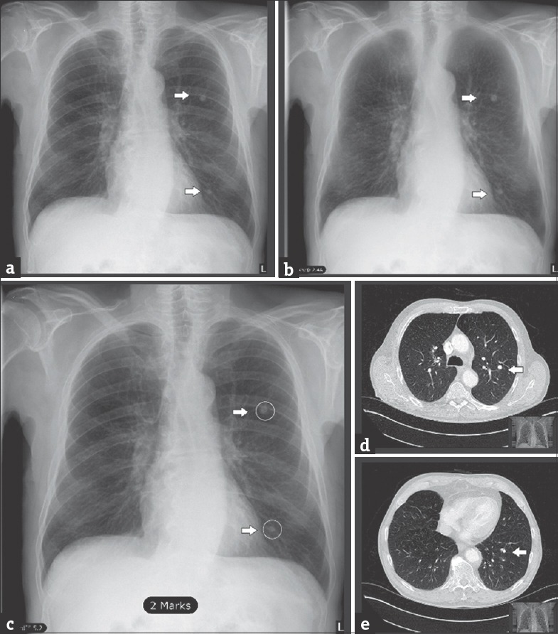
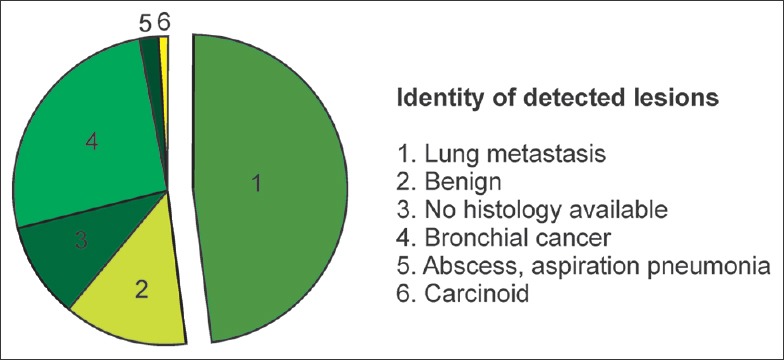
We retrospectively evaluated a dataset of 100 posteroanterior chest radiographs with pulmonary nodular lesions ranging from 5 to 85 mm. All nodules were confirmed with a consecutive computed tomography scan and histologically classified as 75% malignant. The number of detected lesions by observation in unprocessed images was compared to the number and dignity of CAD-detected lesions in bone-suppressed images (BSIs).

RESULTS

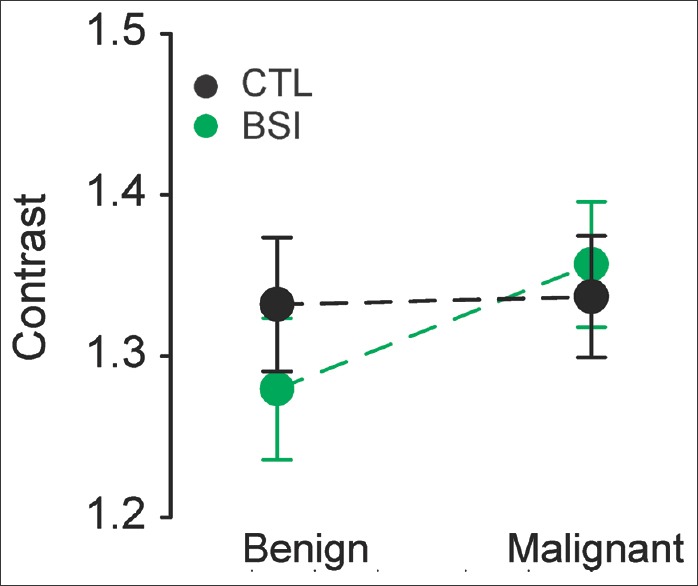
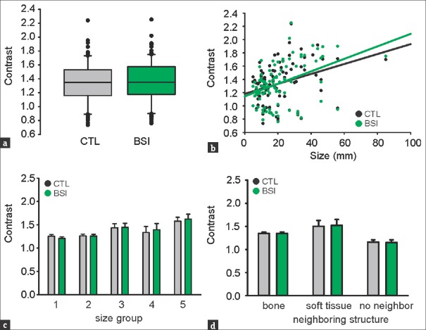
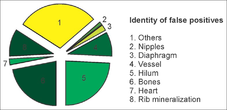
SoftView™ BSI does not affect the objective lesion-to-background contrast. OnGuard™ has a stand-alone sensitivity of 62% and specificity of 58% for nodular lesion detection in chest radiographs. The false positive rate is 0.88/image and the false negative (FN) rate is 0.35/image. From the true positive lesions, 20% were proven benign and 80% were malignant. FN lesions were 47% benign and 53% malignant.

CONCLUSION

We conclude that CAD does not qualify for a stand-alone standard of diagnosis. The use of CAD accompanied with a critical radiological assessment of the software suggested pattern appears more realistic. Accordingly, it is essential to focus on studies assessing the quality-time-cost profile of real-time (as opposed to retrospective) CAD implementation in clinical diagnostics.

摘要

目的

最常用的胸部诊断方法是胸部X光片。一种常见的放射学表现,通常为偶然发现,是肺部结节性病变。从复杂的实质结构中检测小病变是日常临床挑战。在本研究中,我们调查了计算机辅助检测(CAD)软件包SoftView™ 2.4A用于骨抑制的效果以及OnGuard™ 5.2(Riverain Technologies,美国俄亥俄州米亚米斯堡)用于在胸部X光片中自动检测肺结节的效果。

对象与方法

我们回顾性评估了100例后前位胸部X光片的数据集,这些片子上有大小从5到85毫米的肺部结节性病变。所有结节均经连续计算机断层扫描确认,并经组织学分类,75%为恶性。将未处理图像中通过观察检测到的病变数量与骨抑制图像(BSIs)中CAD检测到的病变数量和特征进行比较。

结果

SoftView™ BSI不影响目标病变与背景的对比度。OnGuard™在胸部X光片中检测结节性病变的独立灵敏度为62%,特异性为58%。假阳性率为0.88/图像,假阴性(FN)率为0.35/图像。在真阳性病变中,20%被证实为良性,80%为恶性。FN病变中47%为良性,53%为恶性。

结论

我们得出结论,CAD不符合独立诊断标准。将CAD与对软件提示模式进行的严格放射学评估相结合使用似乎更符合实际。因此,必须专注于评估在临床诊断中实时(与回顾性相对)实施CAD的质量-时间-成本概况的研究。

https://cdn.ncbi.nlm.nih.gov/pmc/blobs/443e/5341301/7f5df8c63b4e/JCIS-7-8-g008.jpg
https://cdn.ncbi.nlm.nih.gov/pmc/blobs/443e/5341301/2af2c3742f1d/JCIS-7-8-g002.jpg
https://cdn.ncbi.nlm.nih.gov/pmc/blobs/443e/5341301/141bd588f95e/JCIS-7-8-g003.jpg
https://cdn.ncbi.nlm.nih.gov/pmc/blobs/443e/5341301/2ed30f8e709d/JCIS-7-8-g005.jpg
https://cdn.ncbi.nlm.nih.gov/pmc/blobs/443e/5341301/0dfe5ebfbfb0/JCIS-7-8-g006.jpg
https://cdn.ncbi.nlm.nih.gov/pmc/blobs/443e/5341301/7f5df8c63b4e/JCIS-7-8-g008.jpg

相似文献

1
Computer-aided Detection Fidelity of Pulmonary Nodules in Chest Radiograph.胸部X线片中肺结节的计算机辅助检测保真度
J Clin Imaging Sci. 2017 Feb 20;7:8. doi: 10.4103/jcis.JCIS_75_16. eCollection 2017.
2
A comparison of computer-aided detection (CAD) effectiveness in pulmonary nodule identification using different methods of bone suppression in chest radiographs.比较不同胸部 X 射线骨抑制方法在肺结节识别中计算机辅助检测(CAD)的有效性。
J Digit Imaging. 2013 Aug;26(4):651-6. doi: 10.1007/s10278-012-9565-4.
3
Computer-aided detection improves detection of pulmonary nodules in chest radiographs beyond the support by bone-suppressed images.计算机辅助检测可提高胸部 X 线摄影中肺结节的检出率,优于骨抑制图像的支持。
Radiology. 2014 Jul;272(1):252-61. doi: 10.1148/radiol.14131315. Epub 2014 Mar 12.
4
Comparison of dual-energy subtraction and electronic bone suppression combined with computer-aided detection on chest radiographs: effect on human observers' performance in nodule detection.双能减影与电子骨抑制技术联合计算机辅助检测在胸部 X 线摄影中的应用比较:对结节检测中观察者性能的影响。
AJR Am J Roentgenol. 2013 May;200(5):1006-13. doi: 10.2214/AJR.12.8877.
5
Evaluation of a real-time interactive pulmonary nodule analysis system on chest digital radiographic images: a prospective study.胸部数字X线摄影图像实时交互式肺结节分析系统的评估:一项前瞻性研究。
Acad Radiol. 2008 May;15(5):571-5. doi: 10.1016/j.acra.2008.01.018.
6
Computer-aided detection of lung cancer on chest radiographs: effect on observer performance.计算机辅助检测胸部 X 射线肺癌:对观察者性能的影响。
Radiology. 2010 Nov;257(2):532-40. doi: 10.1148/radiol.10092437. Epub 2010 Aug 31.
7
Dual energy subtraction digital radiography improves performance of a next generation computer-aided detection program.双能减影数字射线照相术提高了下一代计算机辅助检测程序的性能。
J Thorac Imaging. 2010 Feb;25(1):41-7. doi: 10.1097/RTI.0b013e3181aa34ed.
8
Deep Convolutional Neural Network-based Software Improves Radiologist Detection of Malignant Lung Nodules on Chest Radiographs.基于深度卷积神经网络的软件提高放射科医生在胸部 X 光片上检测恶性肺结节的能力。
Radiology. 2020 Jan;294(1):199-209. doi: 10.1148/radiol.2019182465. Epub 2019 Nov 12.
9
False-positive reduction in computer-aided diagnostic scheme for detecting nodules in chest radiographs by means of massive training artificial neural network.通过大规模训练人工神经网络减少胸部X光片中结节检测的计算机辅助诊断方案中的假阳性。
Acad Radiol. 2005 Feb;12(2):191-201. doi: 10.1016/j.acra.2004.11.017.
10
True detection versus "accidental" detection of small lung cancer by a computer-aided detection (CAD) program on chest radiographs.真阳性检出与计算机辅助检测(CAD)系统在胸部 X 线片上对小肺癌的“偶然”检出的比较。
J Digit Imaging. 2010 Feb;23(1):66-72. doi: 10.1007/s10278-009-9201-0. Epub 2009 May 7.

引用本文的文献

1
Artificial intelligence applications in personalizing lung cancer management: state of the art and future perspectives.人工智能在肺癌个体化管理中的应用:现状与未来展望。
J Thorac Dis. 2024 Oct 31;16(10):7096-7110. doi: 10.21037/jtd-24-244. Epub 2024 Oct 30.
2
Can Artificial Intelligence Replace Humans for Detecting Lung Tumors on Radiographs? An Examination of Resected Malignant Lung Tumors.人工智能能否取代人类在X光片上检测肺肿瘤?对切除的恶性肺肿瘤的研究。
J Pers Med. 2024 Jan 31;14(2):164. doi: 10.3390/jpm14020164.
3
Generative Adversarial Network (Generative Artificial Intelligence) in Pediatric Radiology: A Systematic Review.

本文引用的文献

1
Bone suppression increases the visibility of invasive pulmonary aspergillosis in chest radiographs.骨质抑制可提高胸部X线片上侵袭性肺曲霉病的可见性。
PLoS One. 2014 Oct 3;9(10):e108551. doi: 10.1371/journal.pone.0108551. eCollection 2014.
2
Computer-aided detection improves detection of pulmonary nodules in chest radiographs beyond the support by bone-suppressed images.计算机辅助检测可提高胸部 X 线摄影中肺结节的检出率,优于骨抑制图像的支持。
Radiology. 2014 Jul;272(1):252-61. doi: 10.1148/radiol.14131315. Epub 2014 Mar 12.
3
A comparison of computer-aided detection (CAD) effectiveness in pulmonary nodule identification using different methods of bone suppression in chest radiographs.
生成对抗网络(生成式人工智能)在儿科放射学中的应用:一项系统综述。
Children (Basel). 2023 Aug 10;10(8):1372. doi: 10.3390/children10081372.
4
Review on chest pathogies detection systems using deep learning techniques.基于深度学习技术的胸部疾病检测系统综述。
Artif Intell Rev. 2023 Mar 20:1-47. doi: 10.1007/s10462-023-10457-9.
5
A systematic approach to deep learning-based nodule detection in chest radiographs.基于深度学习的胸部 X 光片结节检测的系统方法。
Sci Rep. 2023 Jun 21;13(1):10120. doi: 10.1038/s41598-023-37270-2.
6
Assessment of the effect of a comprehensive chest radiograph deep learning model on radiologist reports and patient outcomes: a real-world observational study.评估全面胸部 X 光深度学习模型对放射科医生报告和患者结果的影响:一项真实世界观察性研究。
BMJ Open. 2021 Dec 20;11(12):e052902. doi: 10.1136/bmjopen-2021-052902.
7
Current and emerging artificial intelligence applications in chest imaging: a pediatric perspective.当前和新兴的人工智能在胸部成像中的应用:儿科视角。
Pediatr Radiol. 2022 Oct;52(11):2120-2130. doi: 10.1007/s00247-021-05146-0. Epub 2021 Sep 1.
8
Putting artificial intelligence (AI) on the spot: machine learning evaluation of pulmonary nodules.让人工智能(AI)接受检验:肺结节的机器学习评估
J Thorac Dis. 2020 Nov;12(11):6954-6965. doi: 10.21037/jtd-2019-cptn-03.
9
Deep learning-based detection system for multiclass lesions on chest radiographs: comparison with observer readings.基于深度学习的胸片多类病变检测系统:与观察者读数的比较。
Eur Radiol. 2020 Mar;30(3):1359-1368. doi: 10.1007/s00330-019-06532-x. Epub 2019 Nov 20.
10
Deep learning for chest radiograph diagnosis: A retrospective comparison of the CheXNeXt algorithm to practicing radiologists.深度学习在胸片诊断中的应用:CheXNeXt 算法与临床放射科医生的回顾性比较。
PLoS Med. 2018 Nov 20;15(11):e1002686. doi: 10.1371/journal.pmed.1002686. eCollection 2018 Nov.
比较不同胸部 X 射线骨抑制方法在肺结节识别中计算机辅助检测(CAD)的有效性。
J Digit Imaging. 2013 Aug;26(4):651-6. doi: 10.1007/s10278-012-9565-4.
4
Solitary pulmonary nodule: A diagnostic algorithm in the light of current imaging technique.孤立性肺结节:基于当前影像技术的诊断算法
Avicenna J Med. 2011 Oct;1(2):39-51. doi: 10.4103/2231-0770.90915.
5
The effect of computer-aided detection on radiologist performance in the detection of lung cancers previously missed on a chest radiograph.计算机辅助检测对放射科医生检测胸部 X 线片上漏诊肺癌的影响。
J Thorac Imaging. 2013 Jul;28(4):244-52. doi: 10.1097/RTI.0b013e31826c29ec.
6
Computer-aided detection of malignant lung nodules on chest radiographs: effect on observers' performance.计算机辅助检测胸部 X 线片上的恶性肺结节:对观察者性能的影响。
Korean J Radiol. 2012 Sep-Oct;13(5):564-71. doi: 10.3348/kjr.2012.13.5.564. Epub 2012 Aug 28.
7
Improved detection of focal pneumonia by chest radiography with bone suppression imaging.骨抑制成像改善胸部放射摄影对局灶性肺炎的检出。
Eur Radiol. 2012 Dec;22(12):2729-35. doi: 10.1007/s00330-012-2550-y. Epub 2012 Jul 5.
8
Observer training for computer-aided detection of pulmonary nodules in chest radiography.观察者培训在胸部 X 射线摄影中用于计算机辅助检测肺结节。
Eur Radiol. 2012 Aug;22(8):1659-64. doi: 10.1007/s00330-012-2412-7. Epub 2012 Mar 25.
9
Computer-aided detection of small pulmonary nodules in chest radiographs: an observer study.计算机辅助检测胸部 X 线片中的小肺结节:一项观察者研究。
Acad Radiol. 2011 Dec;18(12):1507-14. doi: 10.1016/j.acra.2011.08.008. Epub 2011 Oct 2.
10
Small lung cancers: improved detection by use of bone suppression imaging--comparison with dual-energy subtraction chest radiography.小肺癌:应用骨抑制成像提高检出率——与双能减影胸部 X 射线摄影的比较。
Radiology. 2011 Dec;261(3):937-49. doi: 10.1148/radiol.11110192. Epub 2011 Sep 23.