Boot W, Gawlitta D, Nikkels P G J, Pouran B, van Rijen M H P, Dhert W J A, Vogely H Ch
Department of Orthopaedics, University Medical Center Utrecht, Utrecht, The Netherlands.
Department of Oral and Maxillofacial Surgery & Special Dental Care, University Medical Center Utrecht, Utrecht, The Netherlands.
Clin Orthop Relat Res. 2017 Jul;475(7):1911-1919. doi: 10.1007/s11999-017-5310-0. Epub 2017 Mar 16.
Uncemented orthopaedic implants rely on the bone-implant interface to provide stability, therefore it is essential that a coating does not interfere with the bone-forming processes occurring at the implant interface. In addition, local application of high concentrations of antibiotics for prophylaxis or treatment of infection may be toxic for osteoblasts and could impair bone growth.
QUESTIONS/PURPOSES: In this animal study, we investigated the effect of a commercially available hydrogel, either unloaded or loaded with 2% vancomycin. We asked, does unloaded hydrogel or hydrogel with vancomycin (1) interfere with bone apposition and timing of bone deposition near the implant surface; and (2) induce a local or systemic inflammatory reaction as determined by inflammation around the implant and hematologic parameters.
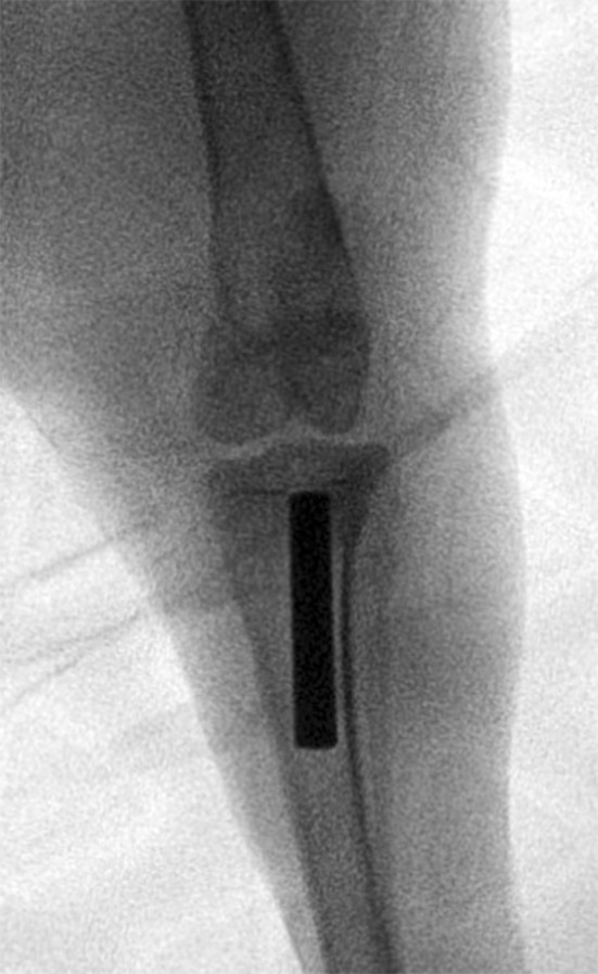
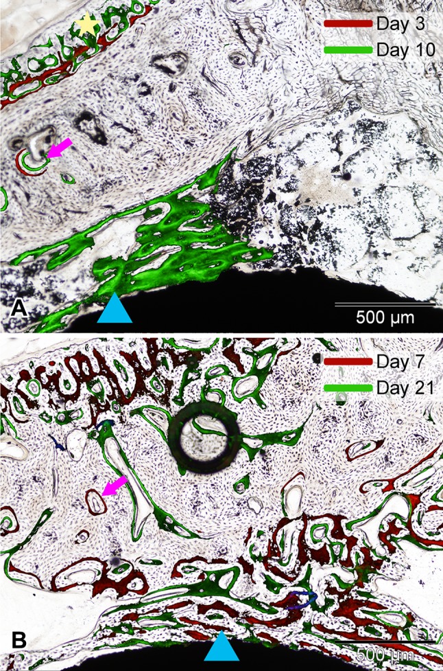
In 18 New Zealand White rabbits, an uncoated titanium rod (n = 6), a rod coated with unloaded hydrogel (n = 6), or a rod coated with 2% vancomycin-loaded hydrogel (n = 6) was implanted in the intramedullary canal of the left tibia. After 28 days, the bone volume fraction near the implant was measured with microCT analysis, inflammation was semiquantitatively scored on histologic sections, and timing of bone apposition was followed by semiquantitative scoring of fluorochrome incorporation on histologic sections. Two observers, blinded to the treatment, scored the sections and reconciled their scores if there was a disagreement. The hematologic inflammatory reaction was analyzed by measuring total and differential leukocyte counts and erythrocyte sedimentation rates in blood. With group sizes of six animals per group, we had 79% power to detect a difference of 25% in histologic scoring for infection and inflammation.
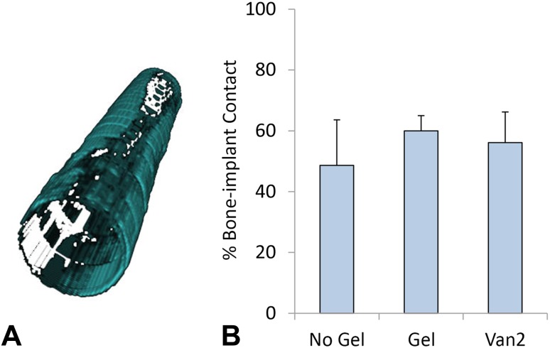
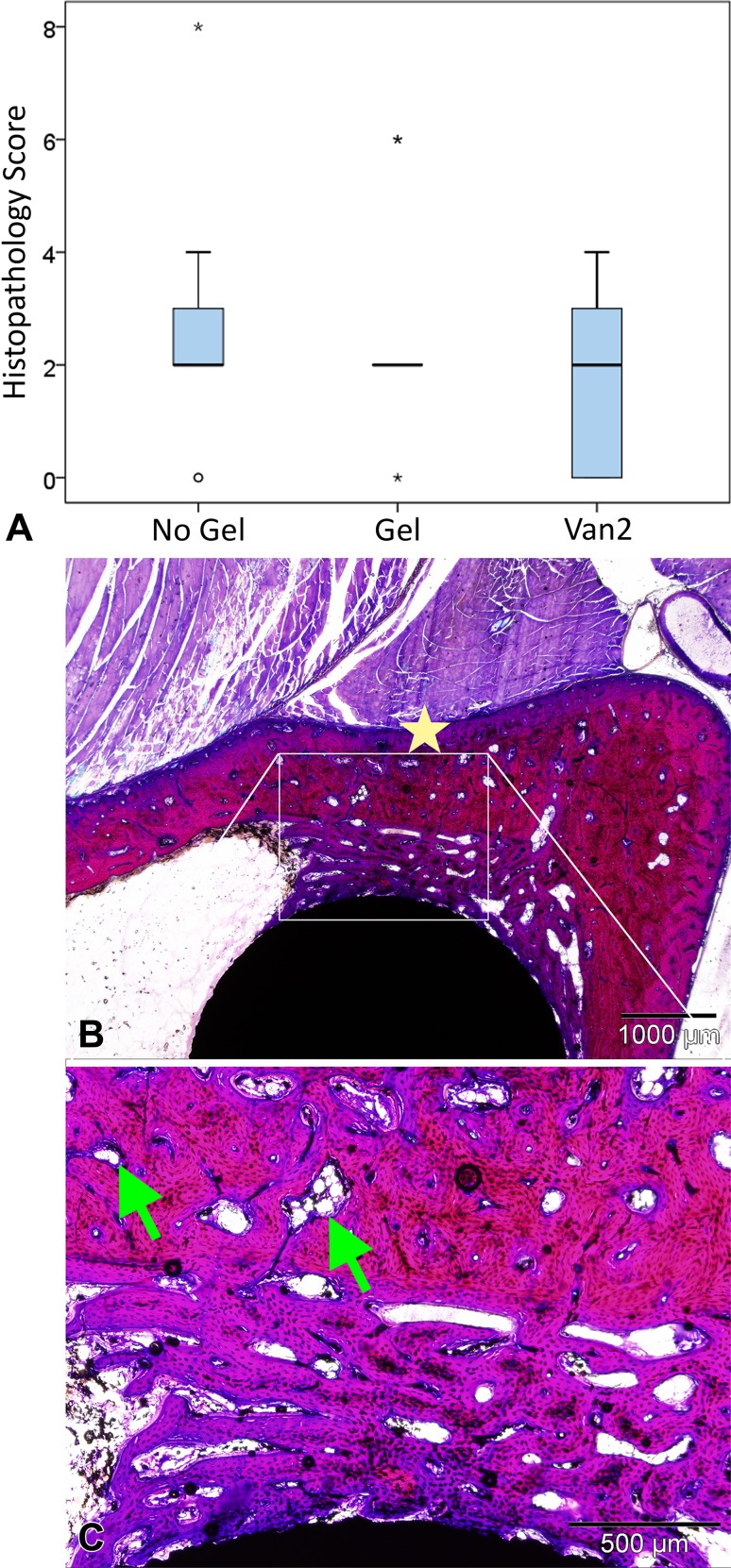
No differences were found in the amount of bone apposition near the implant in the No Gel group (48.65% ± 14.95%) compared with the Gel group (59.97% ± 5.02%; mean difference [MD], 11.32%; 95% CI, -3.89% to 26.53%; p = 0.16) or for the Van2 group (56.12% ± 10.06%; MD, 7.46; 95% CI, -7.75 to 22.67; p = 0.40), with the numbers available. In addition, the scores for timing of bone apposition did not differ between the No Gel group (0.50 ± 0.55) compared with the Gel group (0.33 ± 0.52; MD, -0.17; 95% CI, -0.86 to 0.53; p = 0.78) or the Van2 group (0.83 ± 0.41; MD, 0.33; 95% CI, -0.36 to 1.03; p = 0.42). Furthermore, we detected no differences in the histopathology scores for inflammation in the No Gel group (2.33 ± 1.67) compared with the Gel group (3.17 ± 1.59; MD, 0.83; 95% CI, -0.59 to 2.26; p = 0.31) or to the Van2 group (2.5 ± 1.24; MD, 0.17; 95% CI, -1.26 to 1.59; p = 0.95). Moreover, no differences in total leukocyte count, erythrocyte sedimentation rate, and neutrophil, monocyte, eosinophil, basophil, and lymphocyte counts were present between the No Gel or Van2 groups compared with the Gel control group, with the numbers available.
The hydrogel coated on titanium implants, unloaded or loaded with 2% vancomycin, had no effect on the volume or timing of bone apposition near the implant, and did not induce an inflammatory reaction in vivo, with the numbers available.
Antibiotic-loaded hydrogel may prove to be a valuable option to protect orthopaedic implants from bacterial colonization. Future clinical safety studies will need to provide more evidence that this product does not impair bone formation near the implant and prove the safety of this product.
非骨水泥型骨科植入物依靠骨-植入物界面来提供稳定性,因此涂层不干扰植入物界面处发生的骨形成过程至关重要。此外,局部应用高浓度抗生素预防或治疗感染可能对成骨细胞有毒性,并可能损害骨生长。
问题/目的:在本动物研究中,我们研究了一种市售水凝胶(未负载或负载2%万古霉素)的效果。我们探讨,未负载水凝胶或含万古霉素的水凝胶是否(1)干扰植入物表面附近的骨附着和骨沉积时间;以及(2)如通过植入物周围炎症和血液学参数所确定的那样,诱导局部或全身炎症反应。
在18只新西兰白兔中,将一根未涂层钛棒(n = 6)、一根涂有未负载水凝胶的棒(n = 6)或一根涂有2%万古霉素负载水凝胶的棒(n = 6)植入左胫骨骨髓腔内。28天后,通过显微CT分析测量植入物附近的骨体积分数,在组织学切片上对炎症进行半定量评分,并通过对组织学切片上荧光染料掺入情况进行半定量评分来跟踪骨附着时间。两名对治疗不知情的观察者对切片进行评分,如有分歧则协调他们的评分。通过测量血液中的白细胞总数和分类计数以及红细胞沉降率来分析血液学炎症反应。每组有6只动物,我们有79%的把握检测到感染和炎症组织学评分中25%的差异。
与凝胶组(59.97% ± 5.02%;平均差值[MD],11.32%;95%可信区间,-3.89%至26.53%;p = 0.16)相比,无凝胶组(48.65% ± 14.95%)植入物附近的骨附着量无差异,与Van2组(56.12% ± 10.06%;MD,7.46;95%可信区间,-7.75至22.67;p = 0.40)相比也无差异,现有数据如此。此外,无凝胶组(0.50 ± 0.55)与凝胶组(0.33 ± 0.52;MD,-0.17;95%可信区间,-0.86至0.53;p = 0.78)或Van2组(0.83 ± 0.41;MD,0.33;95%可信区间,-0.36至1.03;p = 0.42)之间的骨附着时间评分无差异。此外,与凝胶组(3.17 ± 1.59;MD,0.83;95%可信区间,-0.59至2.26;p = 0.31)相比,无凝胶组(2.33 ± 1.67)炎症组织病理学评分无差异,与Van2组(2.5 ± 1.24;MD,0.17;95%可信区间,-1.26至1.59;p = 0.95)相比也无差异。此外,与凝胶对照组相比,无凝胶组或Van2组在白细胞总数、红细胞沉降率以及中性粒细胞、单核细胞、嗜酸性粒细胞、嗜碱性粒细胞和淋巴细胞计数方面无差异,现有数据如此。
涂覆在钛植入物上的水凝胶,未负载或负载2%万古霉素,对植入物附近的骨体积或骨附着时间没有影响,并且在现有数据范围内未在体内诱导炎症反应。
负载抗生素的水凝胶可能被证明是保护骨科植入物免受细菌定植的一种有价值的选择。未来的临床安全性研究将需要提供更多证据证明该产品不会损害植入物附近的骨形成并证明该产品的安全性。