Ma Chenming, Chen Na, Mao Yurong, Huang Dongfeng, Song Rong, Li Le
Department of Rehabilitation Medicine, Guangdong Engineering Technology Research Center for Rehabilitation Medicine and Clinical Translation, The First Affiliated Hospital, Sun Yat-sen University, Guangzhou, China; Key Laboratory of Sensing Technology and Biomedical Instrument of Guang Dong Province, School of Engineering, Sun Yat-sen University, Guangzhou, China.
Department of Rehabilitation Medicine, Guangdong Engineering Technology Research Center for Rehabilitation Medicine and Clinical Translation, The First Affiliated Hospital, Sun Yat-sen University , Guangzhou , China.
Front Neurol. 2017 Mar 3;8:70. doi: 10.3389/fneur.2017.00070. eCollection 2017.
This study investigates changes in the neuromuscular activation pattern of the lower limb muscles in stroke survivors when crossing obstacles of three different heights.
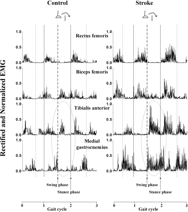
Eight stroke survivors and eight age-, height-, and gender-matched healthy controls were recruited and instructed to cross over obstacles with heights of 10, 20, and 30% leg length. Surface electromyography (EMG) signals were recorded from the rectus femoris (RF), biceps femoris (BF), tibialis anterior (TA), and medial gastrocnemius (MG) of both limbs. Muscle activation signals were normalized to maximum voluntary contraction. Differences between groups and heights were compared using the root mean square of EMG, the cocontraction index of agonist and antagonist muscles, and power spectral analysis based on the mean power frequency (MPF). The correlations between the calculated variables and clinical scales such as Berg Balance Scale and Fugl-Meyer assessment (FMA) were also examined.
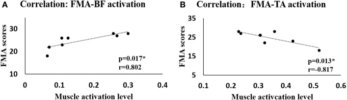
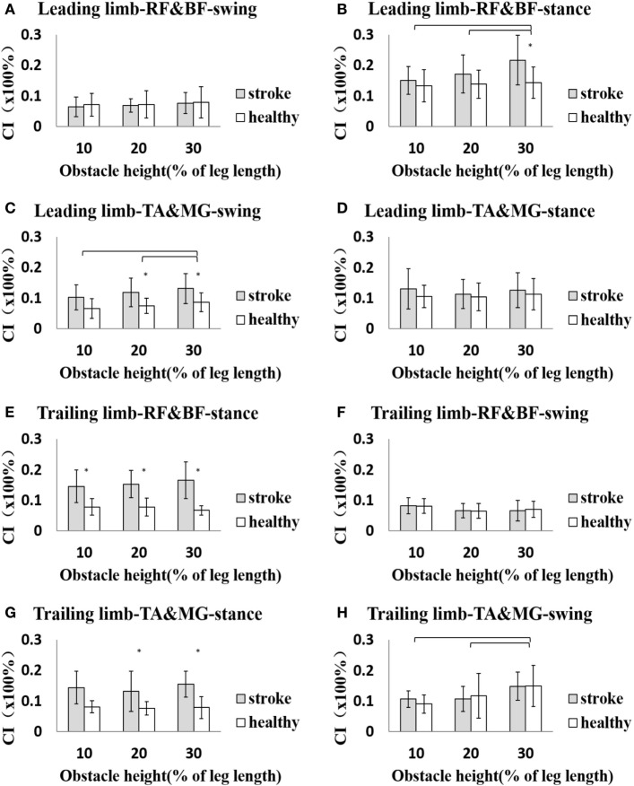
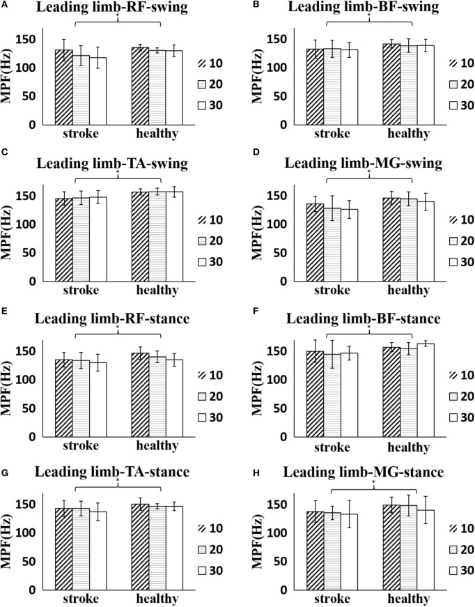
During the leading limb swing phase, the activation levels of all four muscles were greater in the stroke group than the healthy controls ( < 0.05), and the TA showed increased activation level with increasing obstacle height in both groups ( < 0.05). Cocontraction between the TA and MG was higher in the stroke group during the swing phase of the leading limb and between the RF and BF during the stance phase ( < 0.05). Similarly, for the trailing limb, increased cocontractions between the two pairs of agonist and antagonist muscles were found during the stance phase in the stroke group ( < 0.05). During the crossing stride, the frequency analysis showed significantly smaller MPF values in all four lower limb muscles in the leading limb of stroke survivors compared with healthy controls ( < 0.05). Moreover, significant correlations were found between the FMA scores and the BF and TA activations in the leading limb during the swing phase ( < 0.05).
Greater activation levels of the lower limb muscles resulted in higher muscular demands for stroke survivors, which might lead to greater difficulty in maintaining balance. The increased cocontraction during obstacle crossing might be compensation for the affected stability and enable safe crossing for stroke survivors. The reduced MPF in the affected limb of the stroke group might be due to impairments in motor units or other complex neuromuscular alterations.
本研究调查中风幸存者在跨越三种不同高度障碍物时下肢肌肉神经肌肉激活模式的变化。
招募了8名中风幸存者和8名年龄、身高和性别匹配的健康对照者,并指导他们跨越高度分别为腿长10%、20%和30%的障碍物。记录双侧股直肌(RF)、股二头肌(BF)、胫骨前肌(TA)和腓肠肌内侧头(MG)的表面肌电图(EMG)信号。肌肉激活信号以最大自主收缩进行标准化。使用EMG的均方根、 agonist和antagonist肌肉的共同收缩指数以及基于平均功率频率(MPF)的功率谱分析来比较组间和高度之间的差异。还检查了计算变量与临床量表如Berg平衡量表和Fugl-Meyer评估(FMA)之间的相关性。
在领先肢体摆动阶段,中风组所有四块肌肉的激活水平均高于健康对照组(<0.05),并且两组中TA的激活水平均随障碍物高度增加而升高(<0.05)。在领先肢体摆动阶段,中风组TA和MG之间的共同收缩较高,在站立阶段RF和BF之间的共同收缩较高(<0.05)。同样,对于跟随肢体,中风组在站立阶段发现两对agonist和antagonist肌肉之间的共同收缩增加(<0.05)。在跨越步幅期间,频率分析显示中风幸存者领先肢体中所有四块下肢肌肉的MPF值明显小于健康对照组(<0.05)。此外,在摆动阶段领先肢体中,FMA评分与BF和TA激活之间存在显著相关性(<0.05)。
下肢肌肉更高的激活水平导致中风幸存者对肌肉的需求更高,这可能导致维持平衡更加困难。障碍物跨越期间共同收缩的增加可能是对受影响稳定性的补偿,并使中风幸存者能够安全跨越。中风组受影响肢体中MPF的降低可能是由于运动单位受损或其他复杂的神经肌肉改变。