Gardinali Noemi Rovaris, Guimarães Juliana Rodrigues, Melgaço Juliana Gil, Kevorkian Yohan Britto, Bottino Fernanda de Oliveira, Vieira Yasmine Rangel, da Silva Aline Campos de Azevedo, Pinto Douglas Pereira, da Fonseca Laís Bastos, Vilhena Leandro Schiavo, Uiechi Edilson, da Silva Maria Cristina Carlan, Moran Julio, Marchevsky Renato Sérgio, Cruz Oswaldo Gonçalves, Otonel Rodrigo Alejandro Arellano, Alfieri Amauri Alcindo, de Oliveira Jaqueline Mendes, Gaspar Ana Maria Coimbra, Pinto Marcelo Alves
Laboratório de Desenvolvimento Tecnológico em Virologia, Oswaldo Cruz, Instituto Oswaldo Cruz, Fundação Oswaldo Cruz, Rio de Janeiro, Brazil.
Serviço de Equivalência e Farmacocinética -SEFAR, Vice-Presidência de Produção e Inovação em Saúde-VPPIS, Fundação Oswaldo Cruz, Rio de Janeiro, Brazil.
PLoS One. 2017 Mar 22;12(3):e0174070. doi: 10.1371/journal.pone.0174070. eCollection 2017.
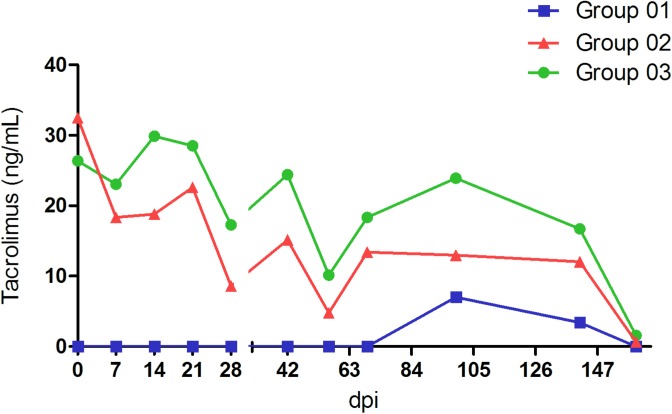
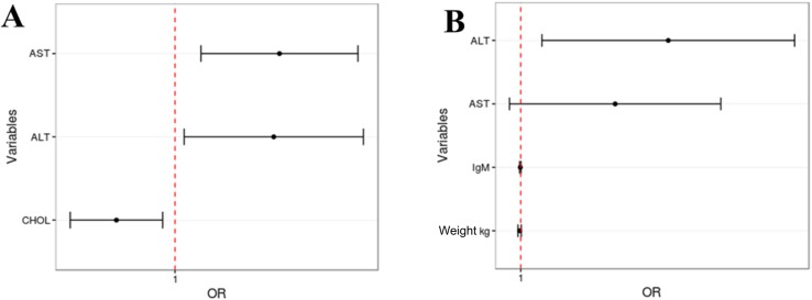
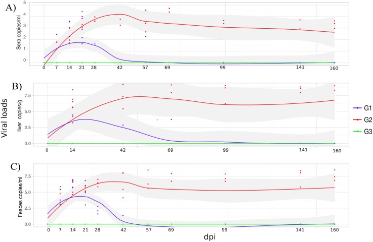
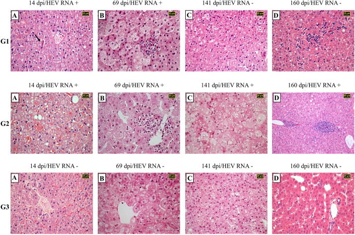
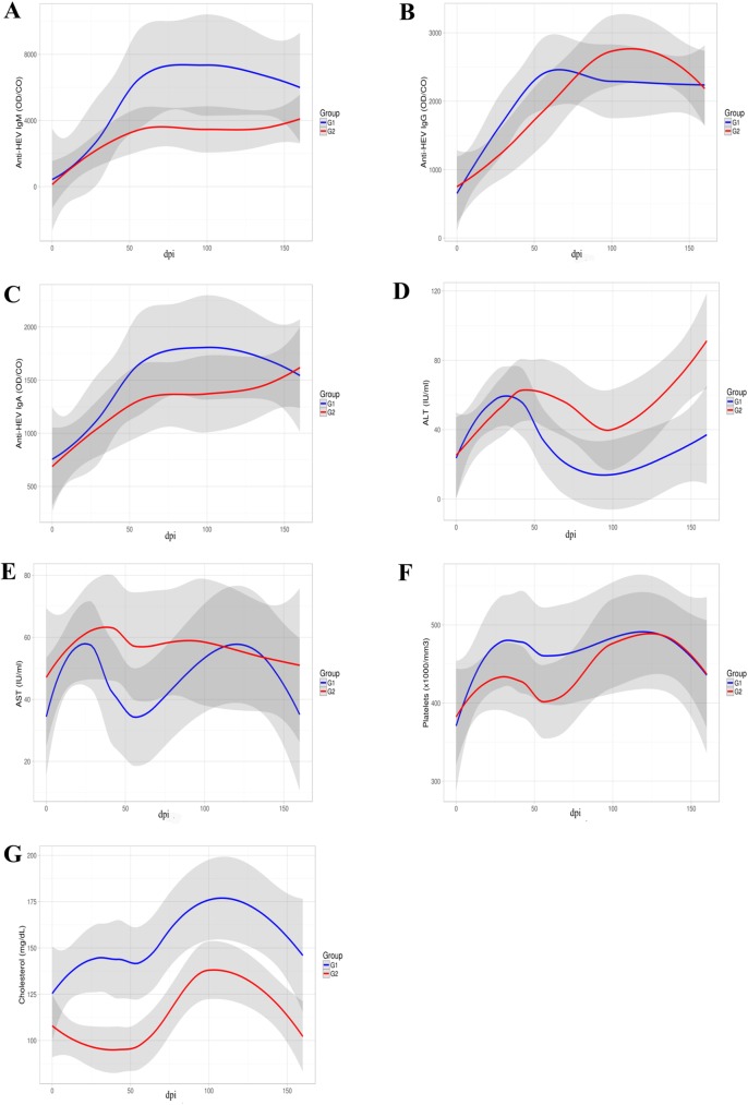
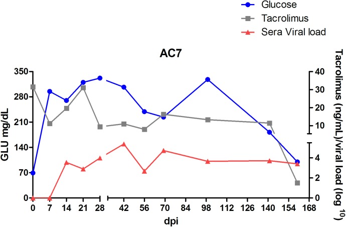
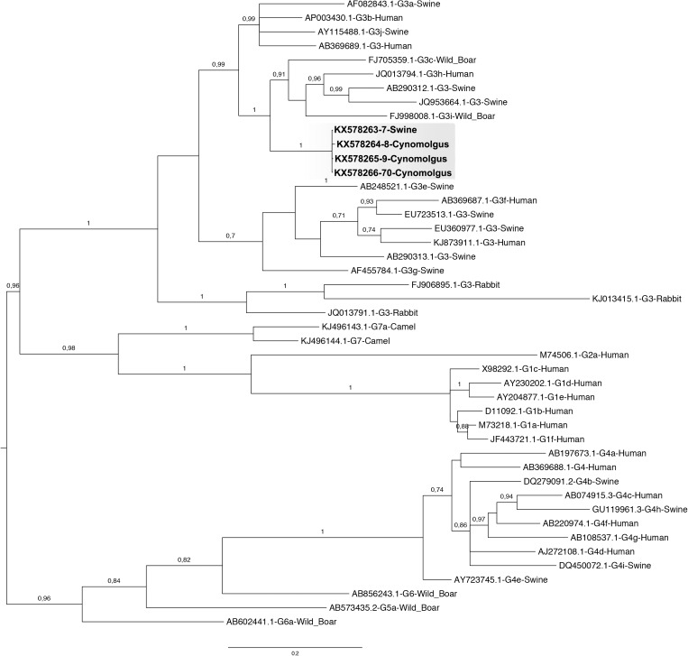
Epidemiological studies found that hepatitis E virus genotype 3 (HEV-3) infection was associated with chronic hepatitis and cirrhosis in immunocompromised patients. Our study aimed to investigate the relationship between the host immunosuppressive status and the occurrence of HEV-related chronic hepatitis. Here we describe a successful experimental study, using cynomolgus monkeys previously treated with tacrolimus, a potent calcineurin inhibitor immunosuppressant, and infected with a Brazilian HEV-3 strain isolated from naturally infected pigs. HEV infected monkeys were followed up during 160 days post infection (dpi) by clinical signs; virological, biochemical and haematological parameters; and liver histopathology. The tacrolimus blood levels were monitored throughout the experiment. Immunosuppression was confirmed by clinical and laboratorial findings, such as: moderate weight loss, alopecia, and herpes virus opportunistic infection. In this study, chronic HEV infection was characterized by the mild increase of liver enzymes serum levels; persistent RNA viremia and viral faecal shedding; and liver histopathology. Three out of four immunosuppressed monkeys showed recurrent HEV RNA detection in liver samples, evident hepatocellular ballooning degeneration, mild to severe macro and microvesicular steatosis (zone 1), scattered hepatocellular apoptosis, and lobular focal inflammation. At 69 dpi, liver biopsies of all infected monkeys revealed evident ballooning degeneration (zone 3), discrete hepatocellular apoptosis, and at most mild portal and intra-acinar focal inflammation. At 160 dpi, the three chronically HEV infected monkeys showed microscopic features (piecemeal necrosis) corresponding to chronic hepatitis in absence of fibrosis and cirrhosis in liver parenchyma. Within 4-months follow up, the tacrolimus-immunosuppressed cynomolgus monkeys infected with a Brazilian swine HEV-3 strain exhibited more severe hepatic lesions progressing to chronic hepatitis without liver fibrosis, similarly as shown in tacrolimus-immunosuppressed solid organ transplant (SOT) recipients. The cause-effect relationship between HEV infection and tacrolimus treatment was confirmed in this experiment.
流行病学研究发现,戊型肝炎病毒3型(HEV-3)感染与免疫功能低下患者的慢性肝炎和肝硬化有关。我们的研究旨在调查宿主免疫抑制状态与戊型肝炎相关慢性肝炎发生之间的关系。在此,我们描述一项成功的实验研究,该研究使用先前用强效钙调神经磷酸酶抑制剂免疫抑制剂他克莫司治疗过的食蟹猴,并感染从自然感染猪中分离出的巴西HEV-3毒株。戊型肝炎感染的猴子在感染后160天(dpi)内通过临床体征、病毒学、生化和血液学参数以及肝脏组织病理学进行随访。在整个实验过程中监测他克莫司的血药浓度。通过临床和实验室检查结果确认免疫抑制,如:中度体重减轻、脱发和疱疹病毒机会性感染。在本研究中,慢性戊型肝炎感染的特征是肝酶血清水平轻度升高、持续性RNA病毒血症和病毒粪便排出,以及肝脏组织病理学变化。四只免疫抑制的猴子中有三只在肝脏样本中反复检测到戊型肝炎病毒RNA,有明显的肝细胞气球样变性、轻度至重度大泡性和小泡性脂肪变性(1区)、散在的肝细胞凋亡以及小叶局灶性炎症。在69 dpi时,所有感染猴子的肝脏活检显示明显的气球样变性(3区)、离散的肝细胞凋亡,以及至多轻度的门管区和腺泡内局灶性炎症。在160 dpi时,三只慢性戊型肝炎感染的猴子显示出与慢性肝炎相对应的微观特征(桥接坏死),肝实质无纤维化和肝硬化。在4个月的随访期内,感染巴西猪戊型肝炎病毒3型毒株的他克莫司免疫抑制食蟹猴表现出更严重的肝脏病变,进展为无肝纤维化的慢性肝炎,这与他克莫司免疫抑制实体器官移植(SOT)受者的情况相似。本实验证实了戊型肝炎感染与他克莫司治疗之间的因果关系。