Ajibade Peter A, Botha Nandipha L
Department of Chemistry, University of Fort Hare, Private Bag X1314, Alice 5700, South Africa.
Nanomaterials (Basel). 2017 Feb 4;7(2):32. doi: 10.3390/nano7020032.
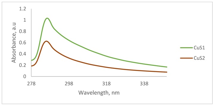
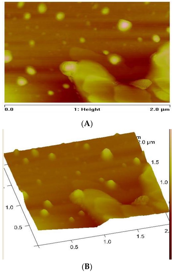
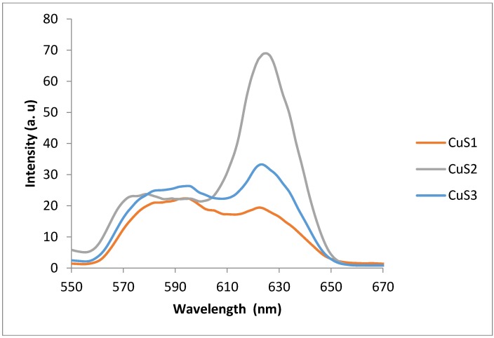
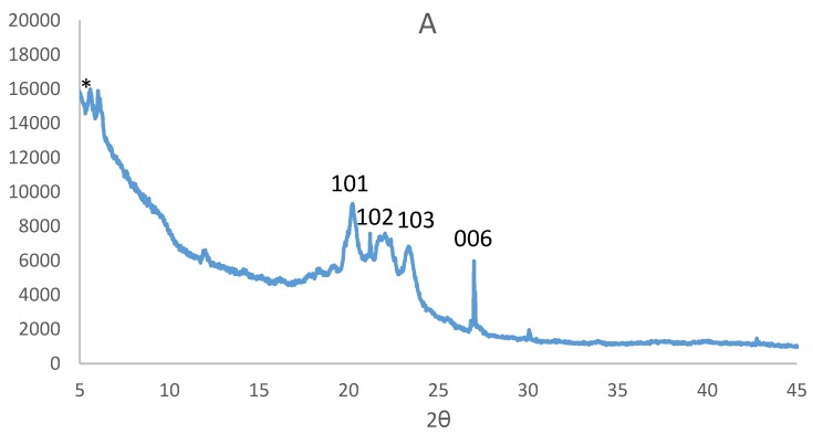
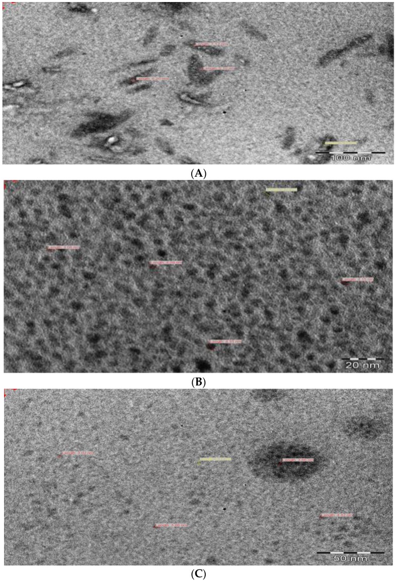
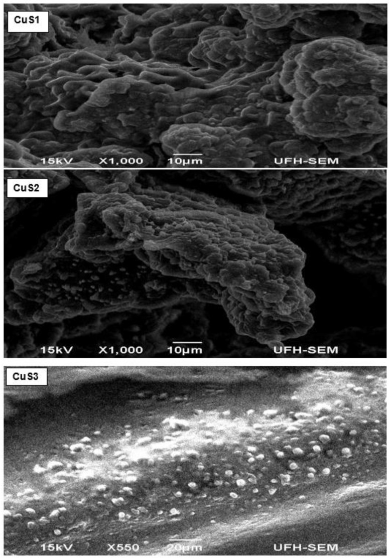
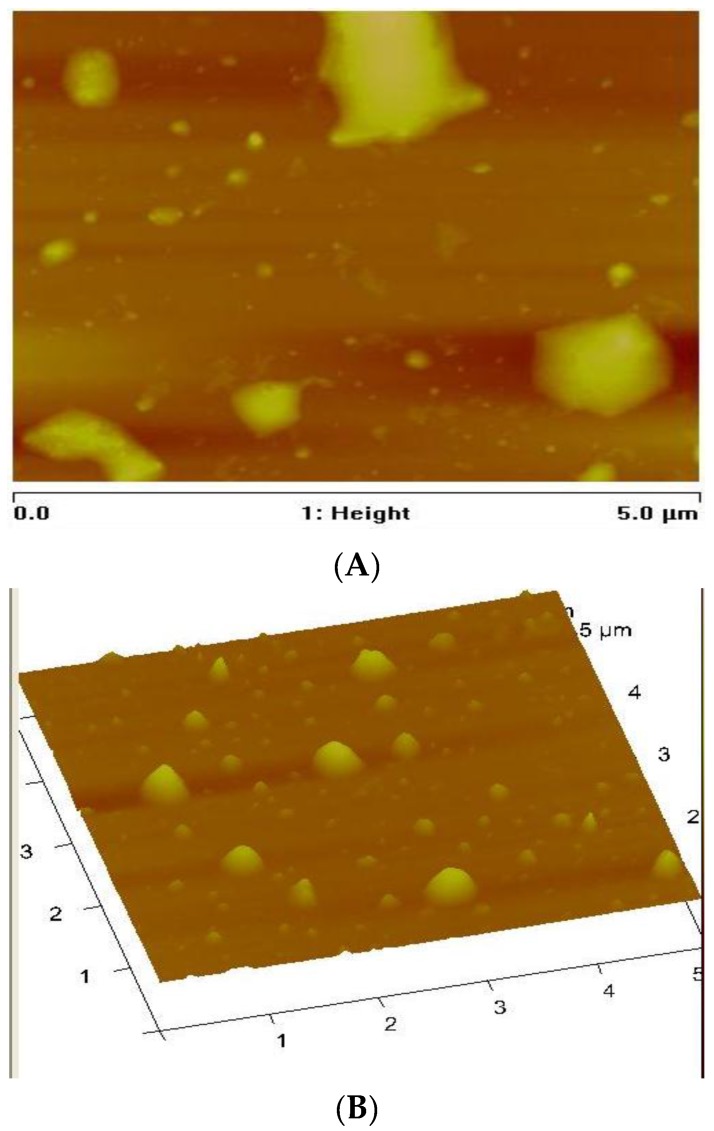
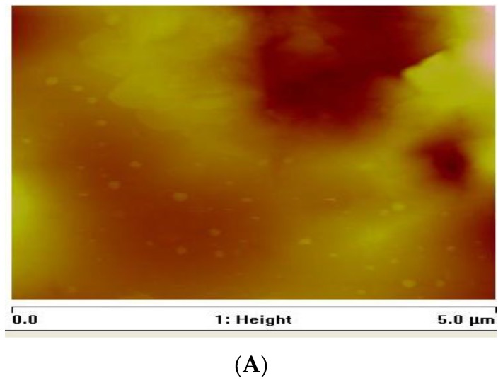
We report the synthesis and structural studies of copper sulfide nanocrystals from copper (II) dithiocarbamate single molecule precursors. The precursors were thermolysed in hexadecylamine (HDA) to prepare HDA-capped CuS nanocrystals. The optical properties of the nanocrystals studied using UV-visible and photoluminescence spectroscopy showed absorption band edges at 287 nm that are blue shifted, and the photoluminescence spectra show emission curves that are red-shifted with respect to the absorption band edges. These shifts are as a result of the small crystallite sizes of the nanoparticles leading to quantum size effects. The structural studies were carried out using powder X-ray diffraction (XRD), transmission electron microscopy (TEM), scanning electron microscopy (SEM), energy dispersive X-ray spectroscopy (EDS), and atomic force microscopy. The XRD patterns indicates that the CuS nanocrystals are in hexagonal covellite crystalline phases with estimated particles sizes of 17.3-18.6 nm. The TEM images showed particles with almost spherical or rod shapes, with average crystallite sizes of 3-9.8 nm. SEM images showed morphology with ball-like microspheres on the surfaces, and EDS spectra confirmed the presence of CuS nanoparticles.
我们报道了由二硫代氨基甲酸盐铜(II)单分子前驱体制备硫化铜纳米晶体的合成及结构研究。将前驱体在十六胺(HDA)中热解以制备HDA包覆的硫化铜纳米晶体。使用紫外可见光谱和光致发光光谱对纳米晶体的光学性质进行研究,结果表明其吸收带边缘在287nm处发生蓝移,并且光致发光光谱显示发射曲线相对于吸收带边缘发生红移。这些位移是由于纳米颗粒的微晶尺寸较小导致量子尺寸效应所致。使用粉末X射线衍射(XRD)、透射电子显微镜(TEM)、扫描电子显微镜(SEM)、能量色散X射线光谱(EDS)和原子力显微镜进行结构研究。XRD图谱表明硫化铜纳米晶体为六方纤锌矿晶相,估计颗粒尺寸为17.3 - 18.6nm。TEM图像显示颗粒几乎为球形或棒状,平均微晶尺寸为3 - 9.8nm。SEM图像显示表面有球状微球的形态,EDS光谱证实了硫化铜纳米颗粒的存在。