Rahni David Ozzie, Toyonaga Takashi, Ohara Yoshiko, Lombardo Francesco, Baba Shinichi, Takihara Hiroshi, Tanaka Shinwa, Kawara Fumiaki, Azuma Takeshi
Brown University/Rhode Island Hospital, Rhode Island Hospital, Providence, United States.
Department of Endoscopy, Kobe University Hospital, Kobe, Japan; Department of Endoscopy, Kishiwada Tokushukai Hospital, Kishiwada, Japan.
Endosc Int Open. 2017 Mar;5(3):E146-E150. doi: 10.1055/s-0042-122965.
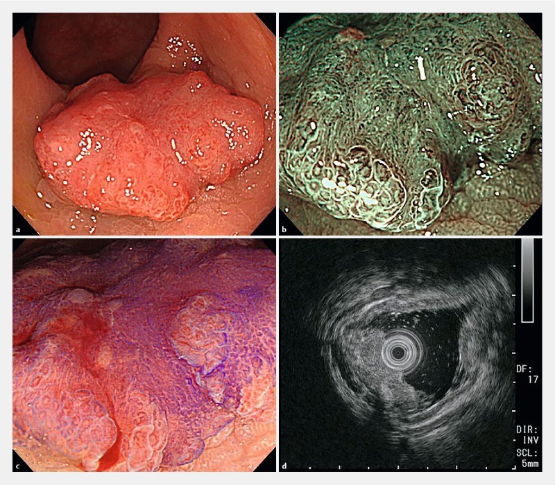
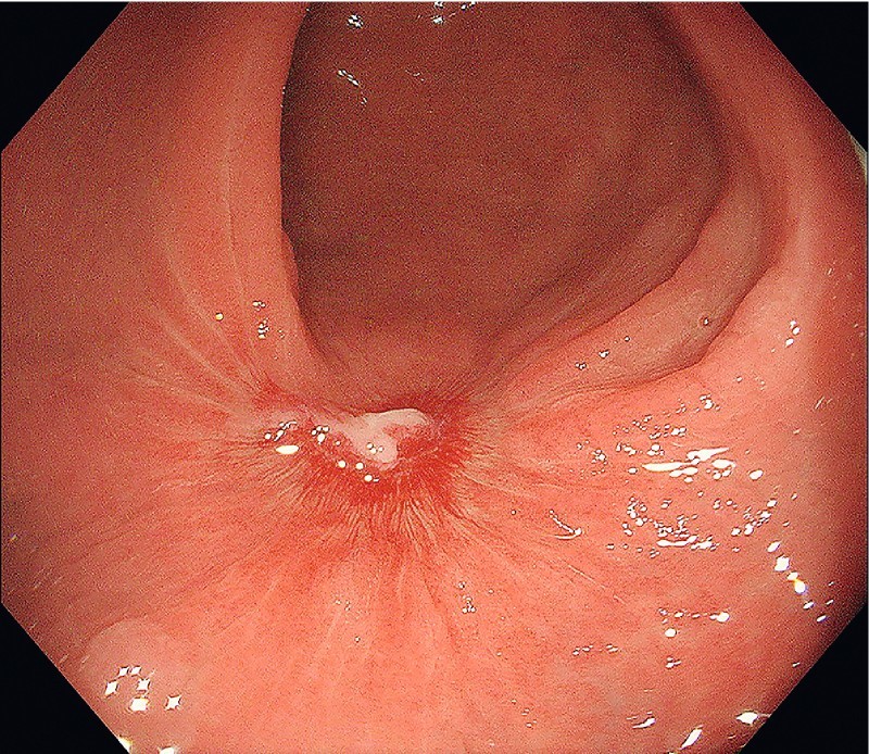
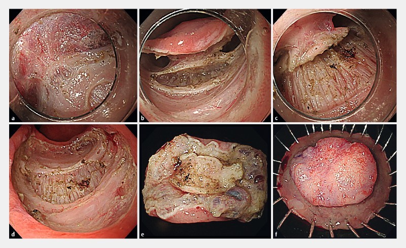
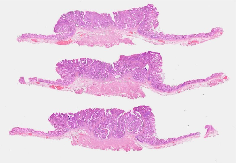
A 54-year-old man was diagnosed with a rectal tumor extending through the submucosal layer. The patient refused surgery and therefore endoscopic submucosal dissection (ESD) was pursued. The lesion exhibited the muscle retraction sign After dissecting circumferentially around the fibrotic area by double tunneling method, a myotomy was performed through the internal circular muscle layer, creating a plane of dissection between the internal circular muscle layer and the external longitudinal muscle layer, and a myectomy was completed. The pathologic specimen verified T1b grade 1 sprouting adenocarcinoma with 4350 µm invasion into the submucosa with negative resection margins.
一名54岁男性被诊断为直肠肿瘤,肿瘤已穿透黏膜下层。患者拒绝手术,因此采用了内镜黏膜下剥离术(ESD)。病变表现出肌肉收缩征。通过双隧道法在纤维化区域周围进行环形剥离后,经内环肌层进行肌切开术,在内环肌层和外纵肌层之间形成一个剥离平面,完成了肌切除术。病理标本证实为T1b 1级芽生性腺癌,侵犯黏膜下层4350 µm,切缘阴性。