Atalay Hasan Anil, Canat H Lütfi, Ülker Volkan, Alkan İlter, Özkuvanci Ünsal, Altunrende Fatih
Department of Urology, Okmeydani Training and Research Hospital, Sisli, Istanbul, Turkey.
Department of Urology, İzmir Tepecik Training and Research Hospital, Izmir, Istanbul, Turkey.
Int Braz J Urol. 2017 May-Jun;43(3):470-475. doi: 10.1590/S1677-5538.IBJU.2016.0441.
To investigate the impact of personalized three dimensional (3D) printed pelvicalyceal system models on patient information before percutaneous nephrolithotripsy surgery.
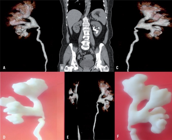
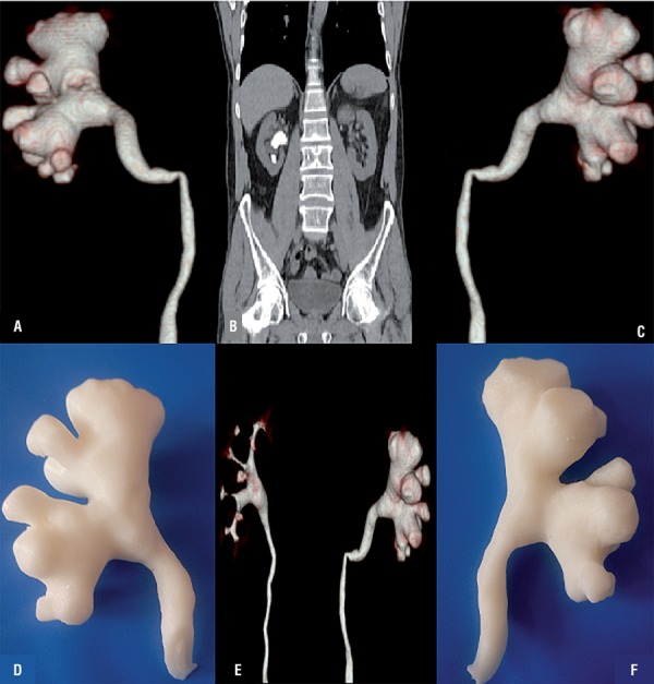
Patients with unilateral complex renal stones with indicatation of percutaneous nephrolithotripsy surgery were selected. Usable data of patients were obtained from CT scans as Digital Imaging and Communications in Medicine (DICOM) format. Mimics software version 16.0 (Materialise, Belgium) was used for segmentation and extraction of pelvicalyceal systems. DICOM format were converted to Stereolithography file format. Finally, fused deposition modeling was used to create plasticine 3D models of pelvicalyceal systems. A questionnaire was designed for patients to assess personalized 3D models effect on patient's understanding their conditions before percutaneous nephrolithotripsy surgery (PCNL). The day before surgery, each patient was seen by a urologist to deliver information about surgery. Questionnaire forms were asked to patients complete before and after presentation of 3D models and the results of the questions were compared.
Five patient's anatomically accurate models of the human renal collecting system were successfully generated. After the 3D printed model presentation, patients demonstrated an improvement in their understanding of basic kidney anatomy by 60% (p=0.017), kidney stone position by 50% (p=0.02), the planned surgical procedure by 60% (p=0.017), and understanding the complications related to the surgery by 64% (p=0.015). In addition, overall satisfaction of conservation improvement was 50% (p=0.02).
Generating kidney models of PCSs using 3D printing technology is feasible, and understandings of the disease and the surgical procedure from patients were well appreciated with this novel technology.
探讨个性化三维(3D)打印肾盂肾盏系统模型对经皮肾镜取石术前患者信息的影响。
选取有经皮肾镜取石术指征的单侧复杂性肾结石患者。患者的可用数据通过CT扫描以医学数字成像和通信(DICOM)格式获取。使用Mimics软件16.0版本(Materialise,比利时)对肾盂肾盏系统进行分割和提取。将DICOM格式转换为立体光刻文件格式。最后,采用熔融沉积建模法制作肾盂肾盏系统的橡皮泥3D模型。设计一份问卷,让患者评估个性化3D模型对经皮肾镜取石术(PCNL)前患者了解自身病情的效果。手术前一天,每位患者由泌尿科医生接诊,以提供有关手术的信息。要求患者在展示3D模型前后填写问卷,并比较问题结果。
成功生成了5例人体肾脏集合系统的解剖学精确模型。3D打印模型展示后,患者对基本肾脏解剖结构的理解提高了60%(p = 0.017),对肾结石位置的理解提高了50%(p = 0.02),对计划手术步骤的理解提高了60%(p = 0.017),对手术相关并发症的理解提高了64%(p = 0.015)。此外,总体沟通改善满意度为50%(p = 0.02)。
利用3D打印技术生成PCS肾脏模型是可行的,这项新技术能让患者对疾病和手术过程有很好的理解。