Department of Anatomy, Catholic Neuroscience Institute, Cell Death Disease Research Center, College of Medicine, The Catholic University of Korea, Seoul, Korea.
Integrative Research Support Center, Laboratory of Electron Microscope, College of Medicine, The Catholic University of Korea, Seoul, Korea.
Sci Rep. 2017 Mar 27;7:45173. doi: 10.1038/srep45173.
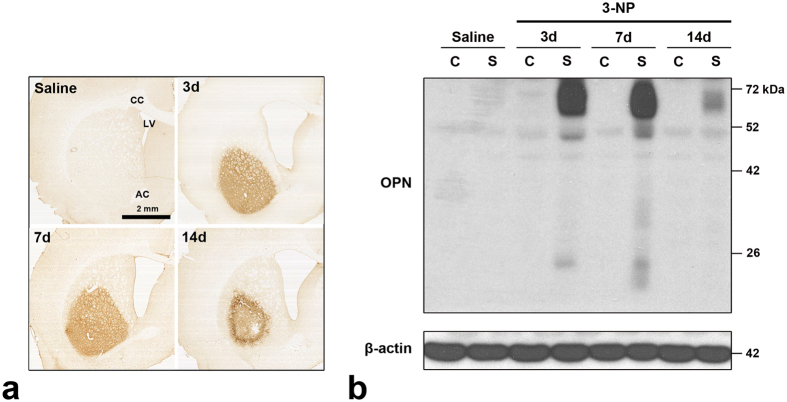
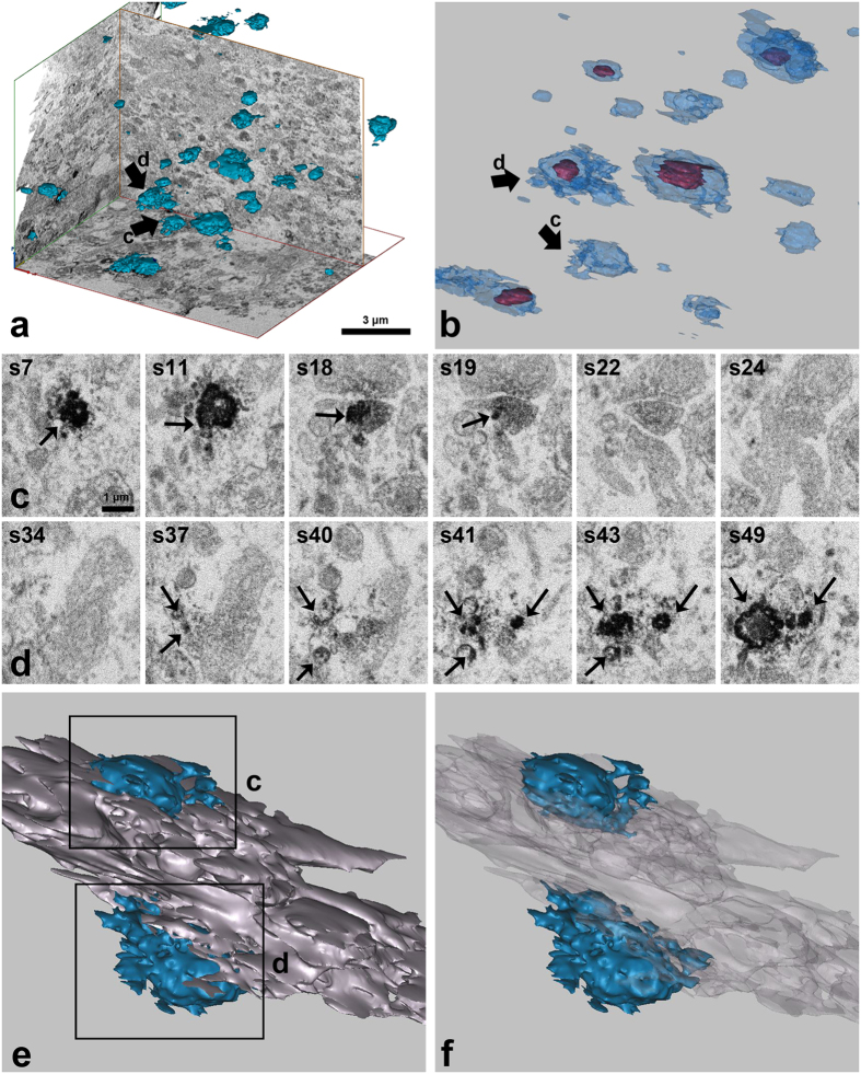
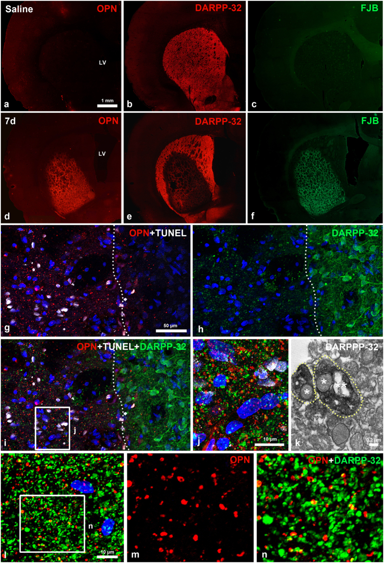
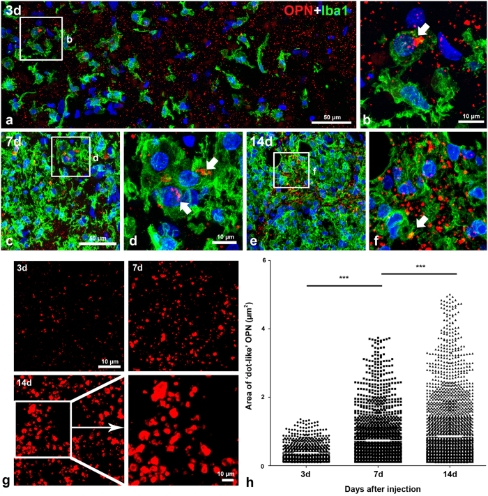
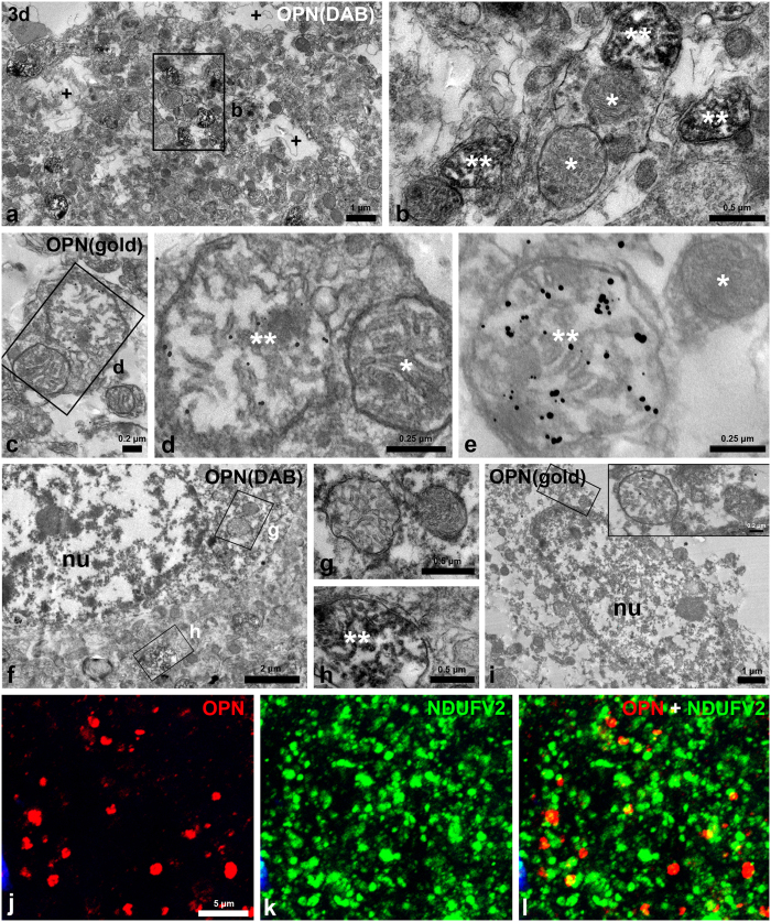
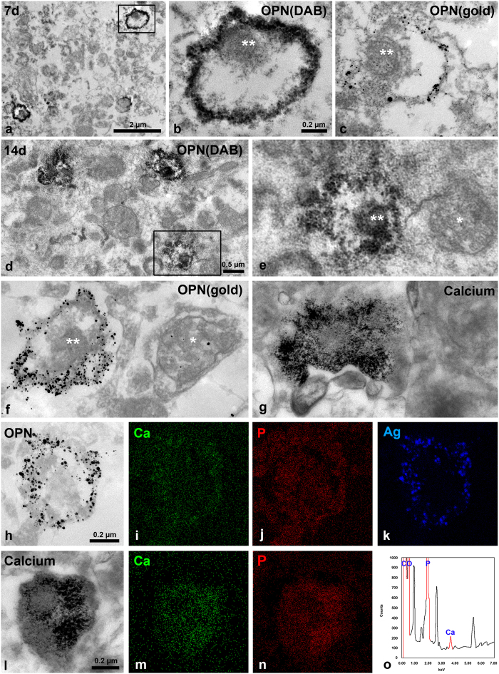
Our aim was to elucidate whether osteopontin (OPN) is involved in the onset of mineralisation and progression of extracellular calcification in striatal lesions due to mitochondrial toxin 3-nitropropionic acid exposure. OPN expression had two different patterns when observed using light microscopy. It was either localised to the Golgi complex in brain macrophages or had a small granular pattern scattered in the affected striatum. OPN labelling tended to increase in number and size over a 2-week period following the lesion. Ultrastructural investigations revealed that OPN is initially localised to degenerating mitochondria within distal dendrites, which were then progressively surrounded by profuse OPN on days 7-14. Electron probe microanalysis of OPN-positive and calcium-fixated neurites indicated that OPN accumulates selectively on the surfaces of degenerating calcifying dendrites, possibly via interactions between OPN and calcium. In addition, 3-dimensional reconstruction of OPN-positive neurites revealed that they are in direct contact with larger OPN-negative degenerating dendrites rather than with fragmented cell debris. Our overall results indicate that OPN expression is likely to correlate with the spatiotemporal progression of calcification in the affected striatum, and raise the possibility that OPN may play an important role in the initiation and progression of microcalcification in response to brain insults.
我们的目的是阐明骨桥蛋白(OPN)是否参与由于暴露于线粒体毒素 3-硝基丙酸而导致纹状体损伤中矿化的开始和细胞外钙化的进展。使用光学显微镜观察时,OPN 表达有两种不同的模式。它要么定位于脑巨噬细胞的高尔基复合体,要么呈小颗粒状散布在受影响的纹状体中。OPN 标记在损伤后 2 周内的数量和大小呈增加趋势。超微结构研究表明,OPN 最初定位于远端树突内的退化线粒体,然后在第 7-14 天逐渐被大量 OPN 包围。对 OPN 阳性和钙固定神经突进行电子探针微分析表明,OPN 选择性地积聚在退化的钙化树突表面,可能是通过 OPN 与钙之间的相互作用。此外,OPN 阳性神经突的三维重建表明,它们与较大的 OPN 阴性退化树突直接接触,而不是与碎片状细胞残骸接触。我们的总体结果表明,OPN 表达可能与受影响纹状体中钙化的时空进展相关,并提出 OPN 可能在脑损伤后微钙化的起始和进展中发挥重要作用的可能性。