Maternal and Fetal Health Centre and Division of Developmental Biology and Medicine, Faculty of Biology, Medicine and Health, University of Manchester, Manchester Academic Health Sciences Centre, St Mary's Hospital, Manchester M13 9WL, UK.
Department of Reproductive Medicine, Old St Mary's Hospital, Central Manchester University Hospitals NHS Foundation Trust, Manchester Academic Health Science Centre, Oxford Road, Manchester M13 9WL, UK.
Cells. 2019 May 9;8(5):432. doi: 10.3390/cells8050432.
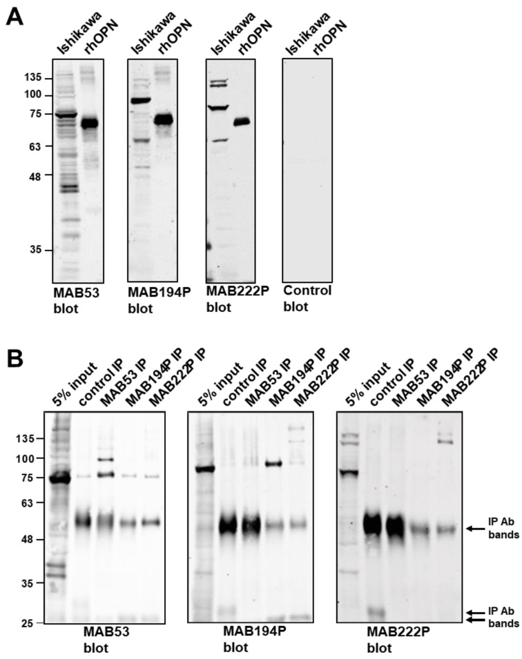
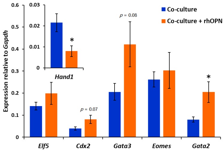
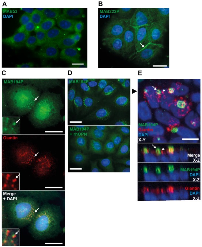
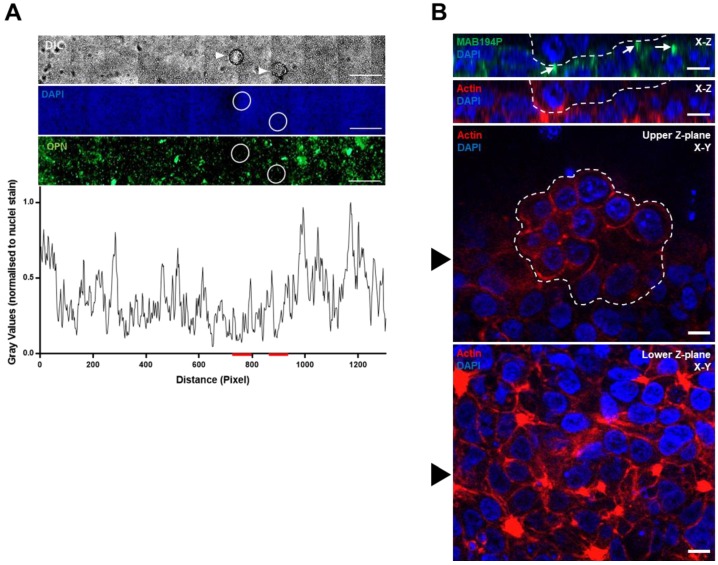
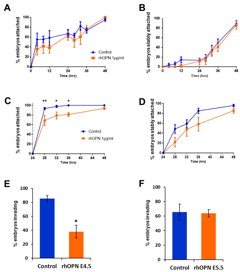
At the onset of pregnancy, embryo implantation is initiated by interactions between the endometrial epithelium and the outer trophectoderm cells of the blastocyst. Osteopontin (OPN) is expressed in the endometrium and is implicated in attachment and signalling roles at the embryo-epithelium interface. We have characterised OPN in the human endometrial epithelial Ishikawa cell line using three different monoclonal antibodies, revealing at least nine distinct molecular weight forms and a novel secretory pathway localisation in the apical domain induced by cell organisation into a confluent epithelial layer. Mouse blastocysts co-cultured with Ishikawa cell layers served to model embryo apposition, attachment and initial invasion at implantation. Exogenous OPN attenuated initial, weak embryo attachment to Ishikawa cells but did not affect the attainment of stable attachment. Notably, exogenous OPN inhibited embryonic invasion of the underlying cell layer, and this corresponded with altered expression of transcription factors associated with differentiation from trophectoderm () to invasive trophoblast giant cells (). These data demonstrate the complexity of endometrial OPN forms and suggest that OPN regulates embryonic invasion at implantation by signalling to the trophectoderm.
在妊娠开始时,胚胎着床是由子宫内膜上皮和囊胚的外层滋养外胚层细胞之间的相互作用启动的。骨桥蛋白(OPN)在子宫内膜中表达,并在胚胎-上皮界面的附着和信号传递作用中起作用。我们使用三种不同的单克隆抗体对人子宫内膜上皮 Ishikawa 细胞系中的 OPN 进行了表征,揭示了至少 9 种不同的分子量形式,以及一种新型的分泌途径定位在细胞组织成致密上皮层时的顶端域中。与 Ishikawa 细胞层共培养的小鼠囊胚用于模拟着床时胚胎的贴附和初始侵袭。外源性 OPN 减弱了初始的、微弱的胚胎附着到 Ishikawa 细胞上,但不影响稳定附着的获得。值得注意的是,外源性 OPN 抑制了胚胎对下一层细胞的侵袭,这与与滋养外胚层()分化为侵袭性滋养层巨细胞()相关的转录因子的表达改变相对应。这些数据表明子宫内膜 OPN 形式的复杂性,并表明 OPN 通过向滋养外胚层发出信号来调节着床时的胚胎侵袭。