Bosch Carles, Martínez Albert, Masachs Nuria, Teixeira Cátia M, Fernaud Isabel, Ulloa Fausto, Pérez-Martínez Esther, Lois Carlos, Comella Joan X, DeFelipe Javier, Merchán-Pérez Angel, Soriano Eduardo
Developmental Neurobiology and Regeneration Unit, Department of Cell Biology and Parc Cientific de Barcelona, University of Barcelona Barcelona, Spain ; Centro de Investigación Biomédica en Red sobre Enfermedades Neurodegenerativas (CIBERNED), Insituto de Salul Carlos III Madrid, Spain ; Institut de Recerca de l'Hospital Universitari de la Vall d'Hebron (VHIR) Barcelona, Spain.
Developmental Neurobiology and Regeneration Unit, Department of Cell Biology and Parc Cientific de Barcelona, University of Barcelona Barcelona, Spain.
Front Neuroanat. 2015 May 21;9:60. doi: 10.3389/fnana.2015.00060. eCollection 2015.
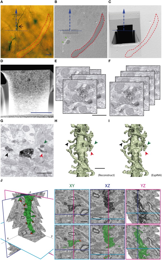
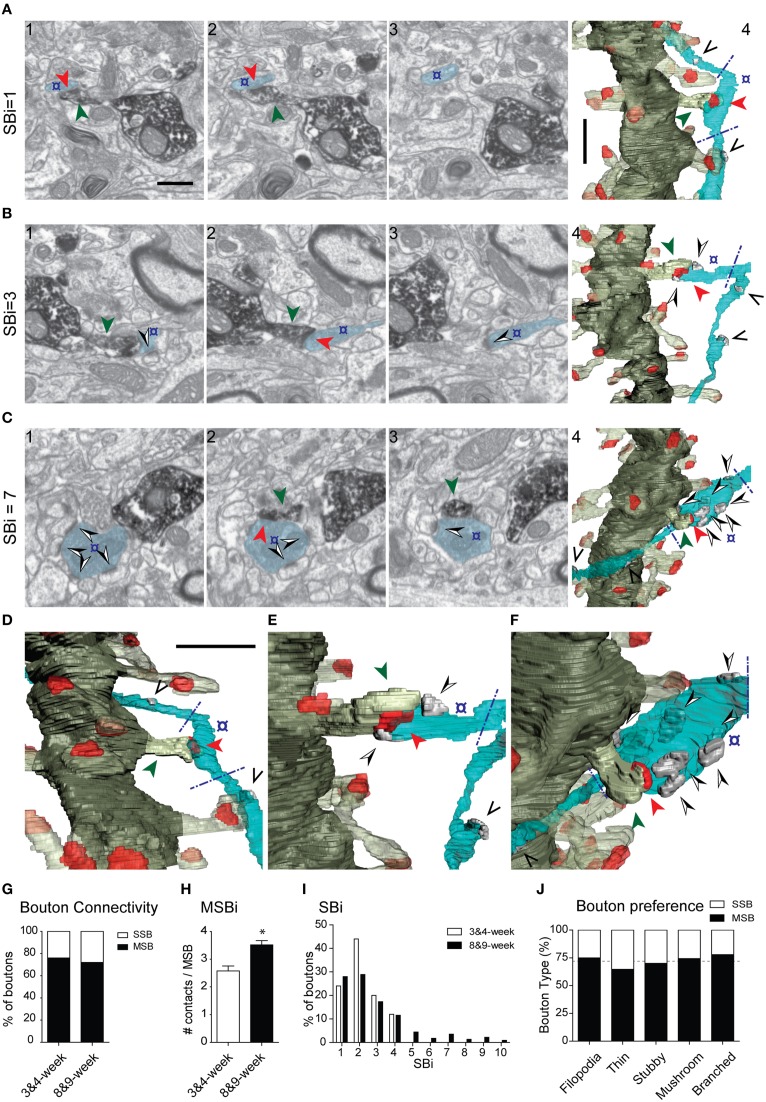
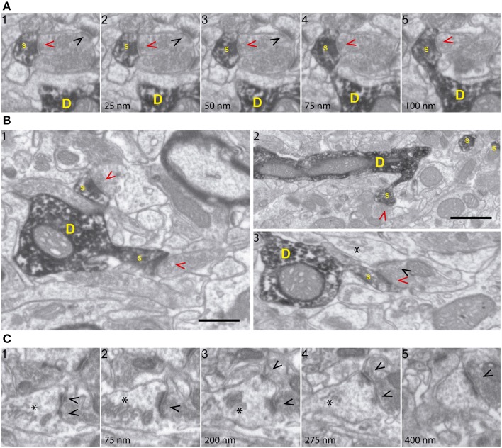
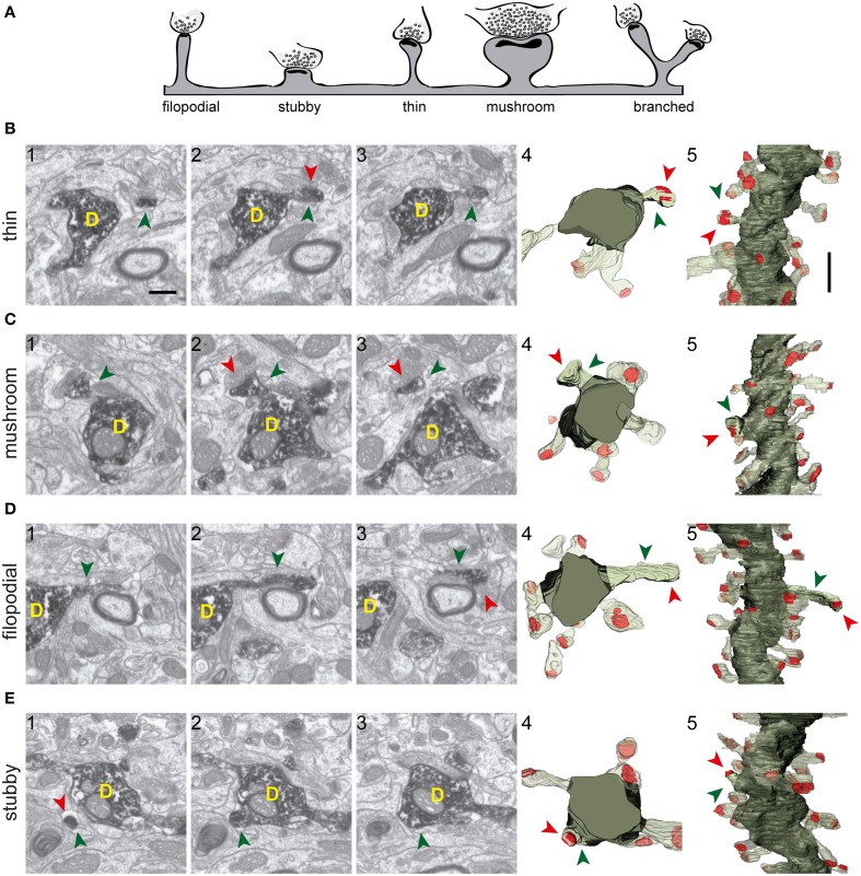
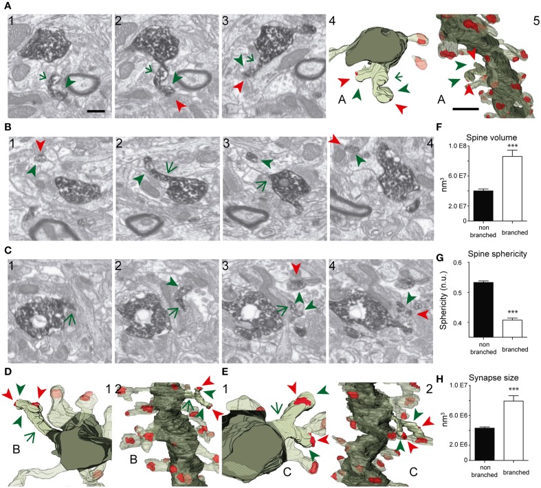
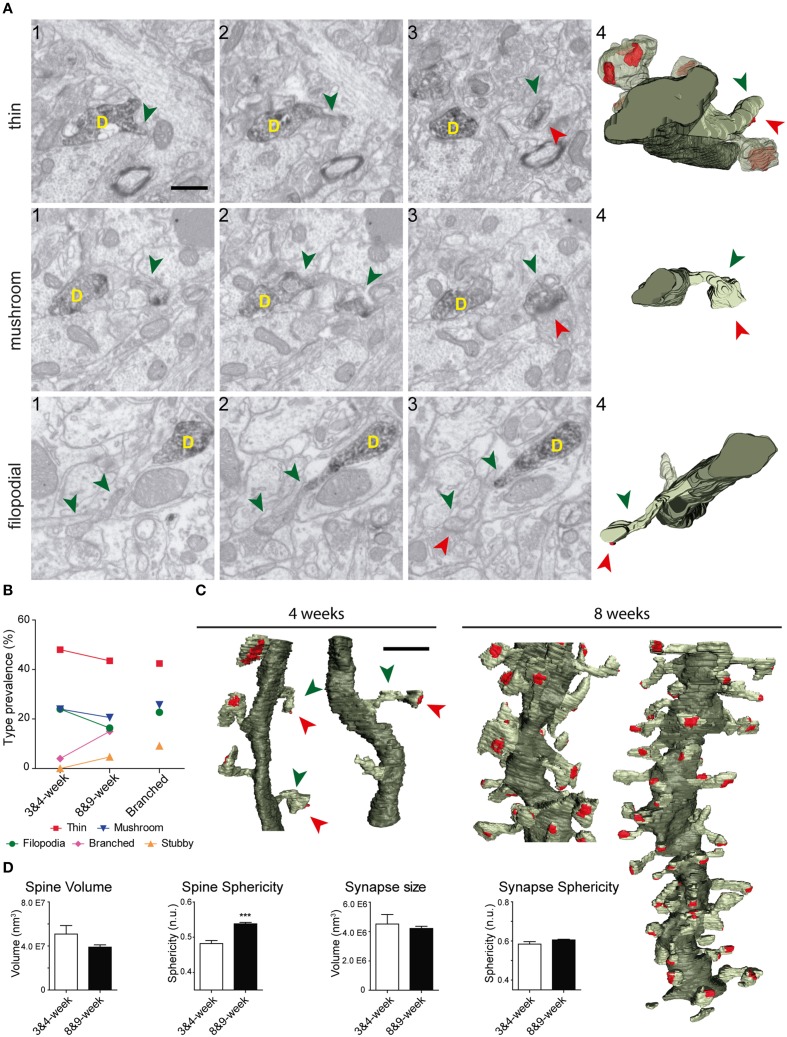
The fine analysis of synaptic contacts is usually performed using transmission electron microscopy (TEM) and its combination with neuronal labeling techniques. However, the complex 3D architecture of neuronal samples calls for their reconstruction from serial sections. Here we show that focused ion beam/scanning electron microscopy (FIB/SEM) allows efficient, complete, and automatic 3D reconstruction of identified dendrites, including their spines and synapses, from GFP/DAB-labeled neurons, with a resolution comparable to that of TEM. We applied this technology to analyze the synaptogenesis of labeled adult-generated granule cells (GCs) in mice. 3D reconstruction of dendritic spines in GCs aged 3-4 and 8-9 weeks revealed two different stages of dendritic spine development and unexpected features of synapse formation, including vacant and branched dendritic spines and presynaptic terminals establishing synapses with up to 10 dendritic spines. Given the reliability, efficiency, and high resolution of FIB/SEM technology and the wide use of DAB in conventional EM, we consider FIB/SEM fundamental for the detailed characterization of identified synaptic contacts in neurons in a high-throughput manner.
通常使用透射电子显微镜(TEM)及其与神经元标记技术的结合来对突触接触进行精细分析。然而,神经元样本复杂的三维结构需要通过连续切片进行重建。在这里,我们展示了聚焦离子束/扫描电子显微镜(FIB/SEM)能够从绿色荧光蛋白/二氨基联苯胺(GFP/DAB)标记的神经元中高效、完整且自动地重建已识别的树突,包括其棘突和突触,分辨率与TEM相当。我们应用这项技术来分析小鼠中标记的成年生成颗粒细胞(GCs)的突触形成。对3 - 4周龄和8 - 9周龄GCs的树突棘进行三维重建,揭示了树突棘发育的两个不同阶段以及突触形成的意外特征,包括空的和分支的树突棘以及与多达10个树突棘建立突触的突触前终末。鉴于FIB/SEM技术的可靠性、效率和高分辨率以及DAB在传统电子显微镜中的广泛应用,我们认为FIB/SEM对于以高通量方式详细表征神经元中已识别的突触接触至关重要。