Cui Yue, Zeng Wenjuan, Yu Jie, Lu Jing, Hu Yuannan, Diao Nan, Liang Bo, Han Ping, Shi Heshui
Department of Radiology, Union Hospital, Tongji Medical College, Huazhong University of Science and Technology, Wuhan, China.
Department of Clinical Laboratory, Union Hospital, Tongji Medical College, Huazhong University of Science and Technology, Wuhan, China.
PLoS One. 2017 Mar 27;12(3):e0174352. doi: 10.1371/journal.pone.0174352. eCollection 2017.
To evaluate the diagnostic performance of left coronary bifurcation angles and plaque characteristics for prediction of coronary stenosis by dual-source CT.
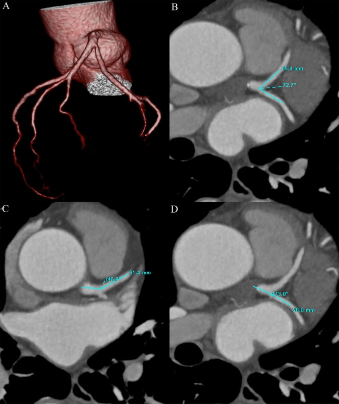
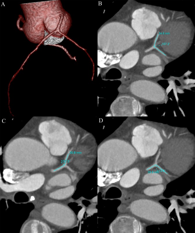
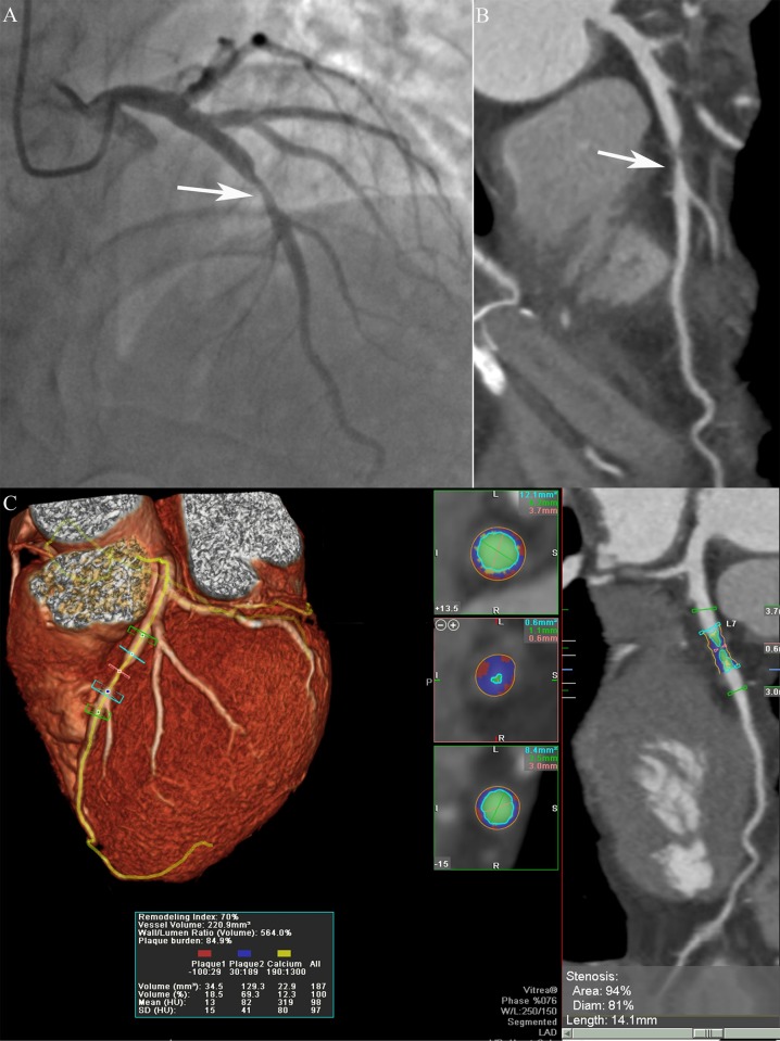
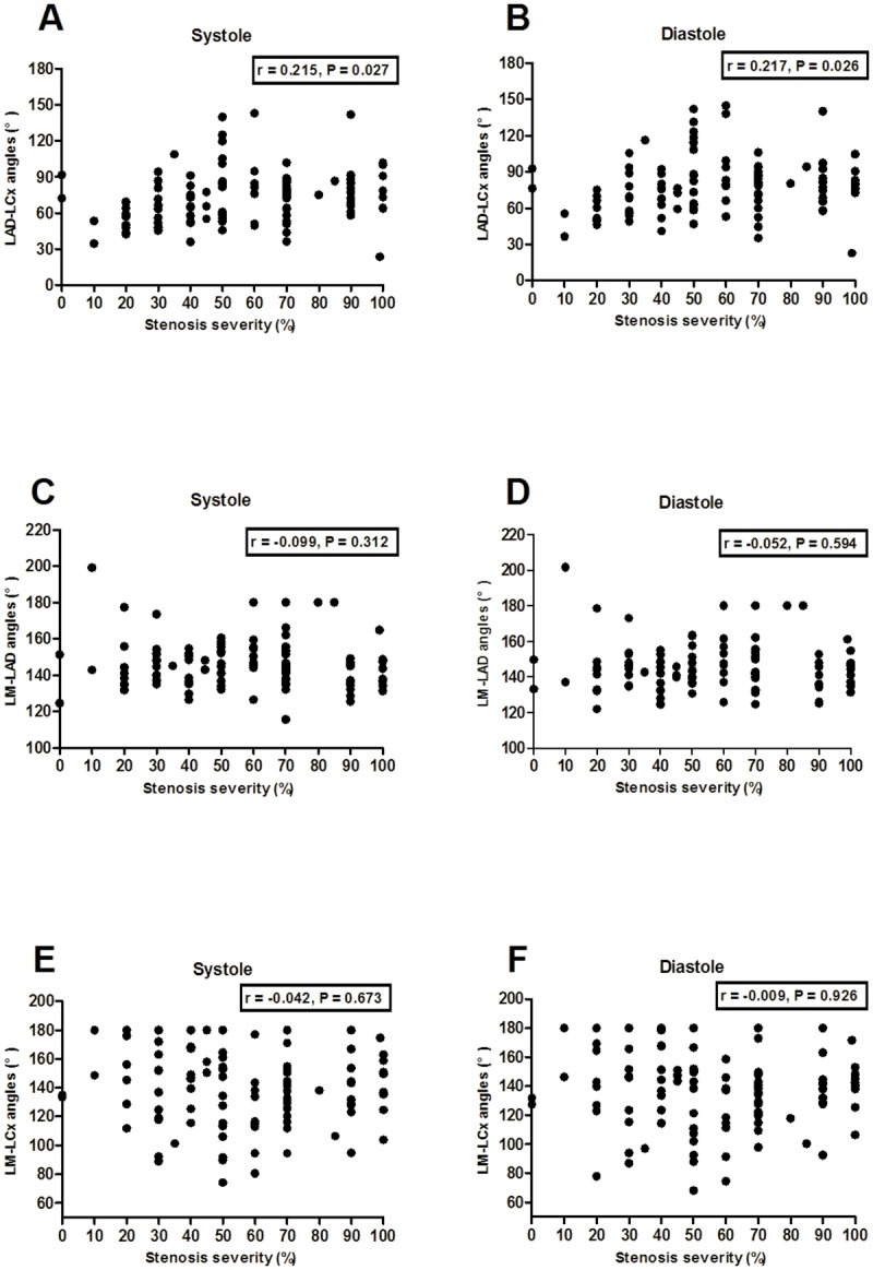
106 patients suspected of coronary artery disease undergoing both coronary computed tomography angiography (CCTA) and invasive coronary angiography (CAG) within three months were included. Left coronary bifurcation angles including the angles between the left anterior descending artery and left circumflex artery (LAD-LCx), left main coronary artery and left anterior descending artery (LM-LAD), left main coronary artery and left circumflex artery (LM-LCx) were measured on CT images. CCTA plaque parameters were calculated by plaque analysis software. Coronary stenosis ≥ 50% by CAG was defined as significant.
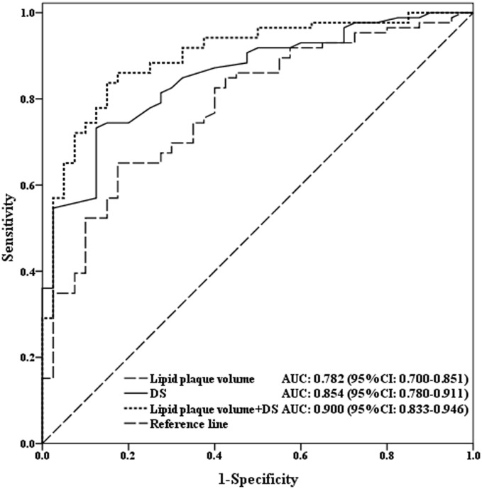
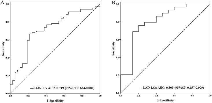
106 patients with 318 left coronary bifurcation angles and 126 vessels were analyzed. The bifurcation angle of LAD-LCx was significantly larger in left coronary stenosis ≥ 50% than stenosis < 50%, and significantly wider in the non-calcified plaque group than calcified. Multivariable analyses showed the bifurcation angle of LAD-LCx was an independent predictor for significant left coronary stenosis (OR = 1.423, P = 0.002). In ROC curve analysis, LAD-LCx predicted significant left coronary stenosis with a sensitivity of 66.7%, specificity of 78.4%, positive predictive value of 85.2% and negative predictive value of 55.8%. The lipid plaque volume improved the diagnostic performance of CCTA diameter stenosis (AUC: 0.854 vs. 0.900, P = 0.045) in significant coronary stenosis.
The bifurcation angle of LAD-LCx could predict significant left coronary stenosis. Wider LAD-LCx is related to non-calcified lesions. Lipid plaque volume could improve the diagnostic performance of CCTA for coronary stenosis prediction.
评估双源CT测量的左冠状动脉分叉角度及斑块特征对预测冠状动脉狭窄的诊断效能。
纳入106例疑似冠心病患者,这些患者在三个月内同时接受了冠状动脉计算机断层扫描血管造影(CCTA)和有创冠状动脉造影(CAG)。在CT图像上测量左冠状动脉分叉角度,包括左前降支与左旋支之间的夹角(LAD-LCx)、左主干与左前降支之间的夹角(LM-LAD)、左主干与左旋支之间的夹角(LM-LCx)。通过斑块分析软件计算CCTA斑块参数。CAG显示冠状动脉狭窄≥50%定义为显著狭窄。
分析了106例患者的318个左冠状动脉分叉角度和126条血管。左冠状动脉狭窄≥50%时,LAD-LCx的分叉角度显著大于狭窄<50%时,且在非钙化斑块组中显著宽于钙化斑块组。多变量分析显示,LAD-LCx的分叉角度是左冠状动脉显著狭窄的独立预测因子(OR = 1.423,P = 0.002)。在ROC曲线分析中,LAD-LCx预测左冠状动脉显著狭窄的敏感性为66.7%,特异性为78.4%,阳性预测值为85.2%,阴性预测值为55.8%。在显著冠状动脉狭窄中,脂质斑块体积提高了CCTA直径狭窄的诊断效能(AUC:0.854对0.900,P = 0.045)。
LAD-LCx的分叉角度可预测左冠状动脉显著狭窄。较宽的LAD-LCx与非钙化病变有关。脂质斑块体积可提高CCTA对冠状动脉狭窄预测的诊断效能。