Iwase Takeshi, Kobayashi Misato, Yamamoto Kentaro, Ra Eimei, Terasaki Hiroko
Department of Ophthalmology, Nagoya University Graduate School of Medicine, Nagoya, Showa-ku, Japan.
PLoS One. 2017 Mar 29;12(3):e0174427. doi: 10.1371/journal.pone.0174427. eCollection 2017.
To investigate ocular blood flow and correlations between ocular blood flow and variables in patients with severe non-proliferative diabetic retinopathy (S-NPDR) following panretinal photocoagulation (PRP).
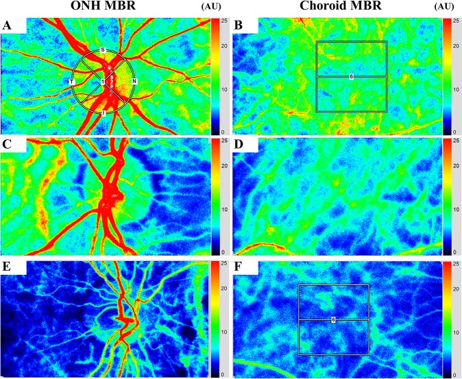
In this retrospective, cross-sectional study, the blood flow on the optic nerve head (ONH) and choroid was assessed with laser speckle flowgraphy (LSFG) using the mean blur rate (MBR) in 76 eyes of 76 patients with S-NPDR who underwent PRP, 39 eyes of 39 patients with S-NPDR who did not undergo PRP, and 71 eyes of 71 normal subjects. The correlation between MBR and variables, including visual acuity (VA) and choroidal area determined by binarization method, was analyzed.
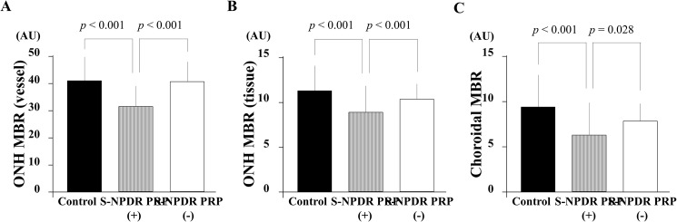
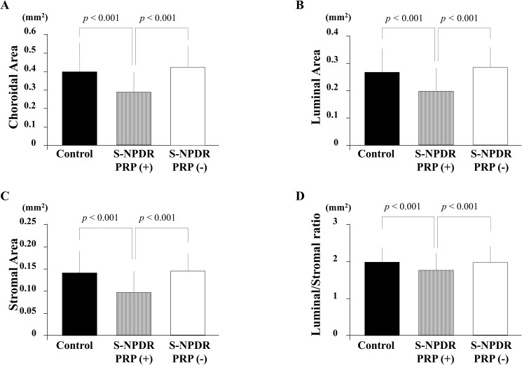
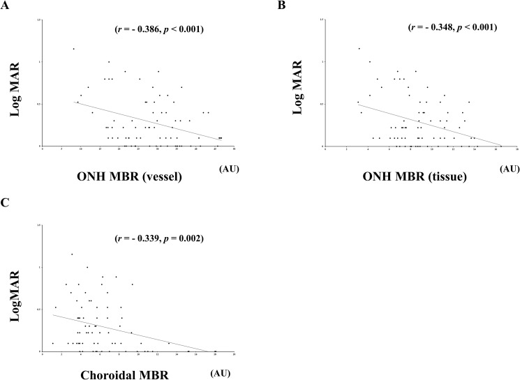
The mean age was 62.9 ± 11.9 years in the S-NPDR with PRP eyes, 55.6 ± 11.4 years in the S-NPDR without PRP eyes, and 60.3 ± 11.1 years in the normal subject eyes. The ONH MBR in vessel and tissue areas and the choroidal MBR were significantly lower in the S-NDR with PRP group than in the other groups (p < 0.001, p < 0.001, and p < 0.001, respectively). The luminal and the stromal areas were significantly smaller in the S-NDR with PRP group than in the other groups (p < 0.001 and p < 0.001, respectively). LogMAR best corrected visual acuity (BCVA) exhibited significant negative correlation with the ONH MBR in vessel (r = -0.386, p < 0.001), tissue (r = -0.348, p < 0.001), and the choroid MBR (r = -0.339, p = 0.002) in the S-NDR with PRP group. Stepwise multiple regression analysis demonstrated that BCVA was a common independent factor associated with the ONH MBR in vessel, tissue, and the choroidal MBR in the S-NDR with PRP group.
ONH and choroid MBR in addition to choroidal component, including the luminal area, were significantly lower in eyes of patients with S-NPDR after PRP compared with no PRP and normal subjects group. This could suggest that the significantly reduced ocular blood flow in PRP-treated S-NPDR eyes correlated with long-term decreased post-PRP luminal area and visual acuity.
研究全视网膜光凝(PRP)后重度非增殖性糖尿病视网膜病变(S-NPDR)患者的眼血流情况以及眼血流与各变量之间的相关性。
在这项回顾性横断面研究中,使用激光散斑血流图(LSFG)通过平均模糊率(MBR)评估了76例接受PRP的S-NPDR患者的76只眼、39例未接受PRP的S-NPDR患者的39只眼以及71例正常受试者的71只眼的视神经乳头(ONH)和脉络膜的血流情况。分析了MBR与包括视力(VA)和通过二值化方法确定的脉络膜面积等变量之间的相关性。
接受PRP的S-NPDR组患者的平均年龄为62.9±11.9岁,未接受PRP的S-NPDR组患者为55.6±11.4岁,正常受试者组为60.3±11.1岁。接受PRP的S-NDR组血管和组织区域的ONH MBR以及脉络膜MBR均显著低于其他组(分别为p<0.001、p<0.001和p<0.001)。接受PRP的S-NDR组的管腔和基质区域显著小于其他组(分别为p<0.001和p<0.001)。在接受PRP的S-NDR组中,LogMAR最佳矫正视力(BCVA)与血管(r=-0.386,p<0.001)、组织(r=-0.348,p<0.001)以及脉络膜MBR(r=-0.339,p=0.002)的ONH MBR呈显著负相关。逐步多元回归分析表明,BCVA是接受PRP的S-NDR组中与血管、组织以及脉络膜MBR的ONH MBR相关的共同独立因素。
与未接受PRP和正常受试者组相比,接受PRP后的S-NPDR患者眼中的ONH和脉络膜MBR以及包括管腔面积在内的脉络膜成分均显著降低。这可能表明,接受PRP治疗的S-NPDR眼中眼血流的显著减少与PRP后长期管腔面积减小和视力下降相关。