• 文献检索
  • 文档翻译
  • 深度研究
  • 学术资讯
  • Suppr Zotero 插件Zotero 插件
  • 邀请有礼
  • 套餐&价格
  • 历史记录
应用&插件
Suppr Zotero 插件Zotero 插件浏览器插件Mac 客户端Windows 客户端微信小程序
定价
高级版会员购买积分包购买API积分包
服务
文献检索文档翻译深度研究API 文档MCP 服务
关于我们
关于 Suppr公司介绍联系我们用户协议隐私条款
关注我们

Suppr 超能文献

核心技术专利:CN118964589B侵权必究
粤ICP备2023148730 号-1Suppr @ 2026

文献检索

告别复杂PubMed语法,用中文像聊天一样搜索,搜遍4000万医学文献。AI智能推荐,让科研检索更轻松。

立即免费搜索

文件翻译

保留排版,准确专业,支持PDF/Word/PPT等文件格式,支持 12+语言互译。

免费翻译文档

深度研究

AI帮你快速写综述,25分钟生成高质量综述,智能提取关键信息,辅助科研写作。

立即免费体验

原代细胞的磷酸化蛋白质组学揭示了卵巢癌中可成药的激酶特征。

Phosphoproteomics of Primary Cells Reveals Druggable Kinase Signatures in Ovarian Cancer.

作者信息

Francavilla Chiara, Lupia Michela, Tsafou Kalliopi, Villa Alessandra, Kowalczyk Katarzyna, Rakownikow Jersie-Christensen Rosa, Bertalot Giovanni, Confalonieri Stefano, Brunak Søren, Jensen Lars J, Cavallaro Ugo, Olsen Jesper V

机构信息

Proteomics Program, Novo Nordisk Foundation Center for Protein Research, Faculty of Health and Medical Sciences, University of Copenhagen, Blegdamsvej 3B, 2200 Copenhagen, Denmark.

Unit of Gynecological Oncology Research, Program of Gynecological Oncology, European Institute of Oncology, Via Ripamonti 435, 20141 Milan, Italy.

出版信息

Cell Rep. 2017 Mar 28;18(13):3242-3256. doi: 10.1016/j.celrep.2017.03.015.

DOI:10.1016/j.celrep.2017.03.015
PMID:28355574
原文链接:https://pmc.ncbi.nlm.nih.gov/articles/PMC5382236/
Abstract

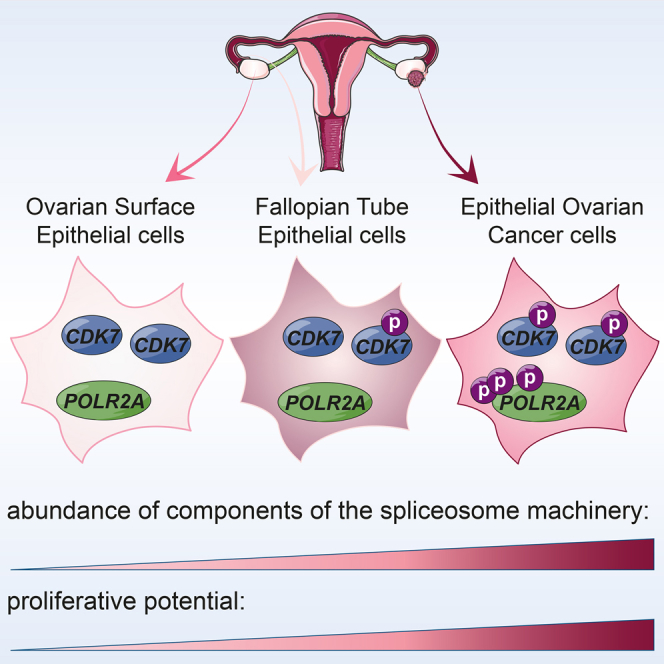
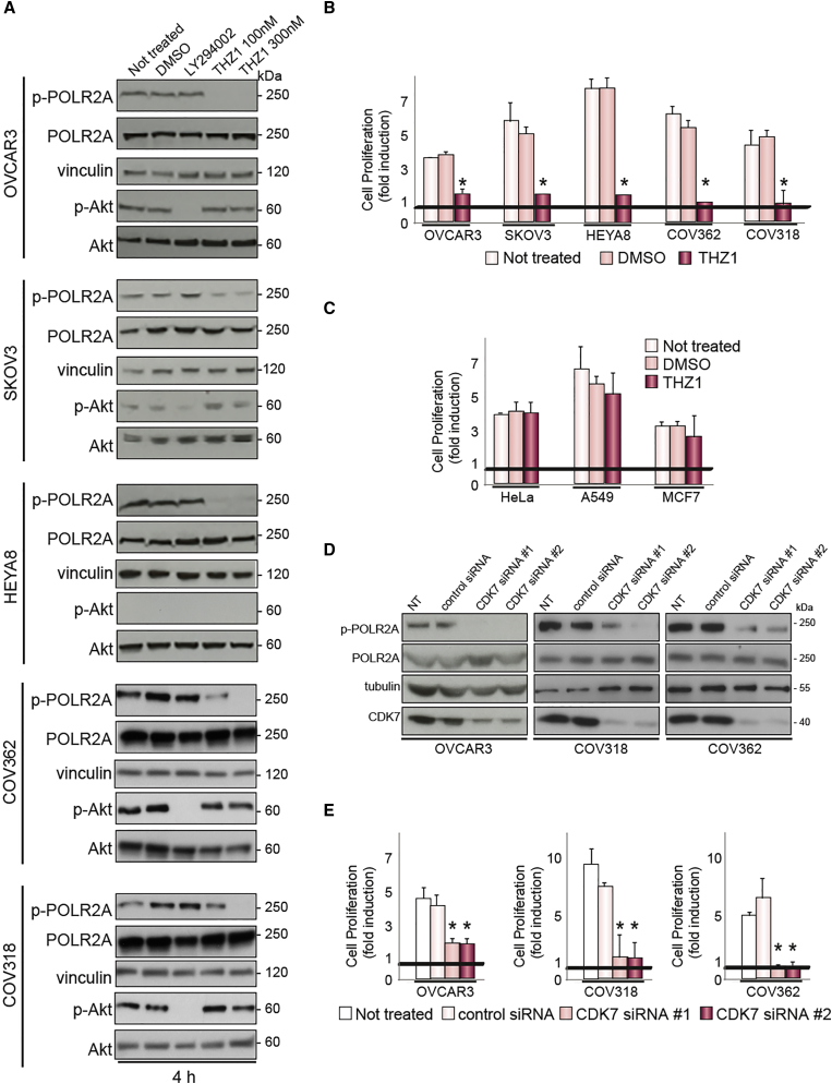
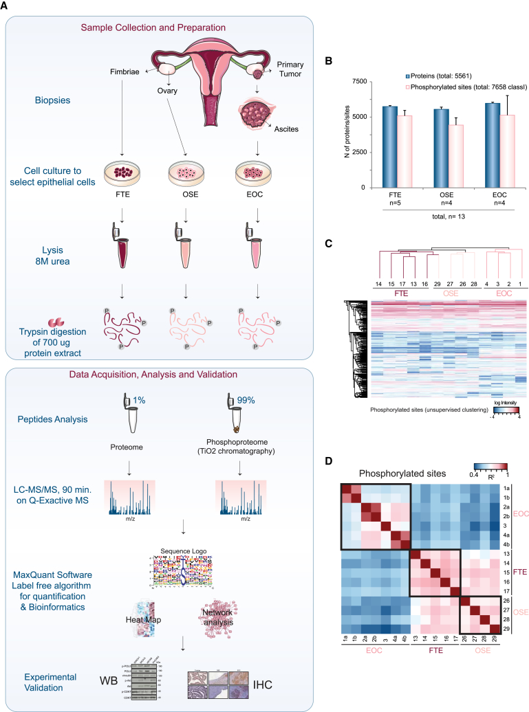
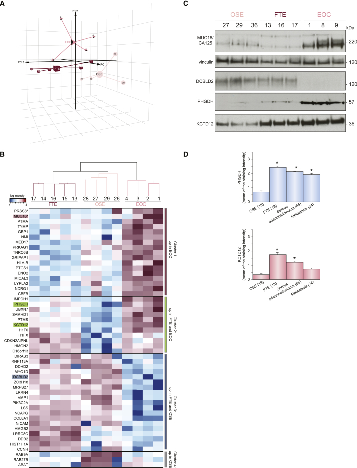
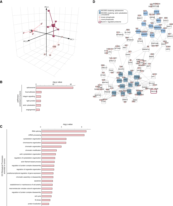
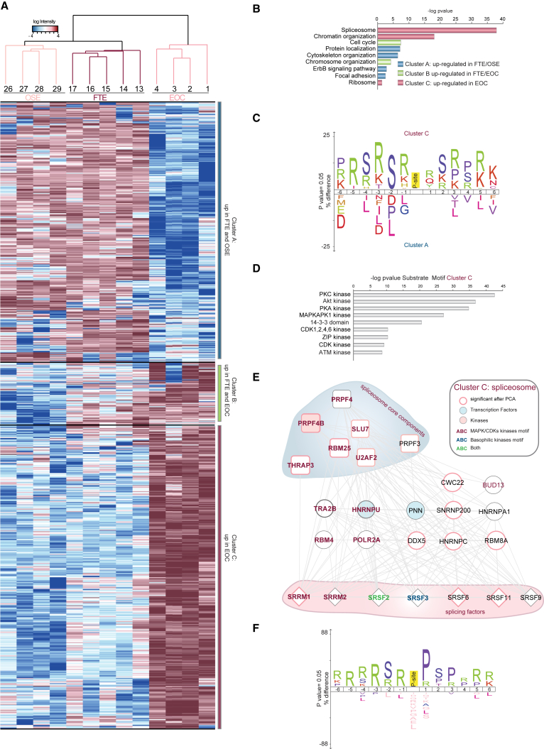
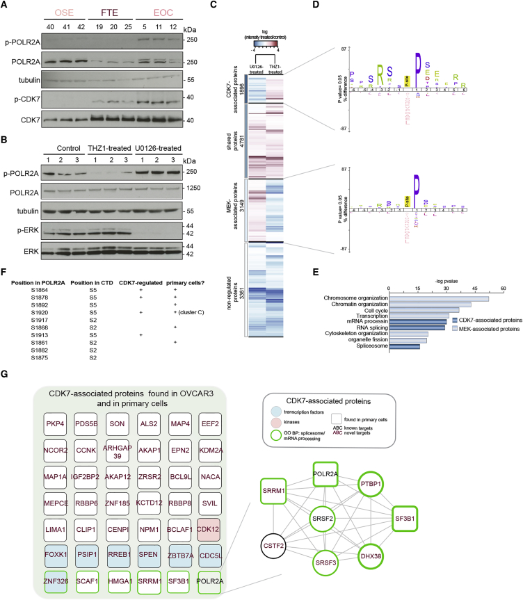
Our understanding of the molecular determinants of cancer is still inadequate because of cancer heterogeneity. Here, using epithelial ovarian cancer (EOC) as a model system, we analyzed a minute amount of patient-derived epithelial cells from either healthy or cancerous tissues by single-shot mass-spectrometry-based phosphoproteomics. Using a multi-disciplinary approach, we demonstrated that primary cells recapitulate tissue complexity and represent a valuable source of differentially expressed proteins and phosphorylation sites that discriminate cancer from healthy cells. Furthermore, we uncovered kinase signatures associated with EOC. In particular, CDK7 targets were characterized in both EOC primary cells and ovarian cancer cell lines. We showed that CDK7 controls cell proliferation and that pharmacological inhibition of CDK7 selectively represses EOC cell proliferation. Our approach defines the molecular landscape of EOC, paving the way for efficient therapeutic approaches for patients. Finally, we highlight the potential of phosphoproteomics to identify clinically relevant and druggable pathways in cancer.

摘要

由于癌症的异质性,我们对癌症分子决定因素的理解仍然不足。在这里,我们以上皮性卵巢癌(EOC)为模型系统,通过基于单次质谱的磷酸化蛋白质组学分析了来自健康或癌组织的微量患者来源的上皮细胞。使用多学科方法,我们证明原代细胞概括了组织复杂性,并且是区分癌症与健康细胞的差异表达蛋白质和磷酸化位点的宝贵来源。此外,我们发现了与EOC相关的激酶特征。特别是,在EOC原代细胞和卵巢癌细胞系中都对CDK7靶点进行了表征。我们表明CDK7控制细胞增殖,并且对CDK7的药理学抑制选择性地抑制EOC细胞增殖。我们的方法定义了EOC的分子格局,为患者的有效治疗方法铺平了道路。最后,我们强调了磷酸化蛋白质组学在识别癌症中临床相关且可药物化途径方面的潜力。

https://cdn.ncbi.nlm.nih.gov/pmc/blobs/fa12/5382236/5387c04f8b29/gr6.jpg
https://cdn.ncbi.nlm.nih.gov/pmc/blobs/fa12/5382236/5d9ac3bcd7d1/fx1.jpg
https://cdn.ncbi.nlm.nih.gov/pmc/blobs/fa12/5382236/87f68d4ec1aa/gr1.jpg
https://cdn.ncbi.nlm.nih.gov/pmc/blobs/fa12/5382236/425acd78f573/gr2.jpg
https://cdn.ncbi.nlm.nih.gov/pmc/blobs/fa12/5382236/3f1d57af4828/gr3.jpg
https://cdn.ncbi.nlm.nih.gov/pmc/blobs/fa12/5382236/e9a0a2f7882e/gr4.jpg
https://cdn.ncbi.nlm.nih.gov/pmc/blobs/fa12/5382236/408f852d4fee/gr5.jpg
https://cdn.ncbi.nlm.nih.gov/pmc/blobs/fa12/5382236/5387c04f8b29/gr6.jpg

相似文献

1
Phosphoproteomics of Primary Cells Reveals Druggable Kinase Signatures in Ovarian Cancer.原代细胞的磷酸化蛋白质组学揭示了卵巢癌中可成药的激酶特征。
Cell Rep. 2017 Mar 28;18(13):3242-3256. doi: 10.1016/j.celrep.2017.03.015.
2
Genetic determinants of FOXM1 overexpression in epithelial ovarian cancer and functional contribution to cell cycle progression.上皮性卵巢癌中FOXM1过表达的遗传决定因素及其对细胞周期进程的功能作用。
Oncotarget. 2015 Sep 29;6(29):27613-27. doi: 10.18632/oncotarget.4546.
3
Discovery of serum biomarkers implicated in the onset and progression of serous ovarian cancer in a rat model using iTRAQ technique.采用 iTRAQ 技术在大鼠模型中发现与浆液性卵巢癌发生和进展相关的血清生物标志物。
Eur J Obstet Gynecol Reprod Biol. 2012 Nov;165(1):96-103. doi: 10.1016/j.ejogrb.2012.06.031. Epub 2012 Jul 18.
4
Molecular subtyping of cancer and nomination of kinase candidates for inhibition with phosphoproteomics: Reanalysis of CPTAC ovarian cancer.癌症的分子亚型分类和磷酸蛋白质组学抑制激酶候选物的提名:CPTAC 卵巢癌的再分析。
EBioMedicine. 2019 Feb;40:305-317. doi: 10.1016/j.ebiom.2018.12.039. Epub 2018 Dec 26.
5
Pharmacological Inhibition of p38 MAPK by SB203580 Increases Resistance to Carboplatin in A2780cp Cells and Promotes Growth in Primary Ovarian Cancer Cells.SB203580 通过抑制 p38 MAPK 增加 A2780cp 细胞对卡铂的耐药性并促进原发性卵巢癌细胞的生长。
Int J Mol Sci. 2018 Jul 26;19(8):2184. doi: 10.3390/ijms19082184.
6
Overexpression of trophoblast cell surface antigen 2 as an independent marker for a poor prognosis and as a potential therapeutic target in epithelial ovarian carcinoma.滋养层细胞表面抗原2的过表达作为上皮性卵巢癌预后不良的独立标志物及潜在治疗靶点。
Int J Exp Pathol. 2016 Apr;97(2):150-8. doi: 10.1111/iep.12174. Epub 2016 Apr 29.
7
LSR Antibody Therapy Inhibits Ovarian Epithelial Tumor Growth by Inhibiting Lipid Uptake.LSR 抗体疗法通过抑制脂质摄取抑制卵巢上皮性肿瘤生长。
Cancer Res. 2018 Jan 15;78(2):516-527. doi: 10.1158/0008-5472.CAN-17-0910. Epub 2017 Nov 29.
8
Novel expression of CD11b in epithelial ovarian cancer: Potential therapeutic target.CD11b 在卵巢上皮性癌中的新型表达:潜在的治疗靶点。
Gynecol Oncol. 2018 Mar;148(3):567-575. doi: 10.1016/j.ygyno.2017.12.018. Epub 2018 Jan 10.
9
Proteomic analysis of ovarian cancer proximal fluids: validation of elevated peroxiredoxin 1 in patient peripheral circulation.卵巢癌近端体液的蛋白质组学分析:患者外周循环中过氧化物酶 1 升高的验证。
PLoS One. 2011;6(9):e25056. doi: 10.1371/journal.pone.0025056. Epub 2011 Sep 30.
10
MicroRNA-145 targets TRIM2 and exerts tumor-suppressing functions in epithelial ovarian cancer.微小RNA-145靶向TRIM2并在上皮性卵巢癌中发挥肿瘤抑制功能。
Gynecol Oncol. 2015 Dec;139(3):513-9. doi: 10.1016/j.ygyno.2015.10.008. Epub 2015 Oct 16.

引用本文的文献

1
Phosphoproteomics Reveals L1CAM-Associated Signaling Networks in High-Grade Serous Ovarian Carcinoma: Implications for Radioresistance and Tumorigenesis.磷酸化蛋白质组学揭示高级别浆液性卵巢癌中与L1细胞粘附分子相关的信号网络:对放射抗性和肿瘤发生的影响
Int J Mol Sci. 2025 May 10;26(10):4585. doi: 10.3390/ijms26104585.
2
Navitoclax, a Bcl-2/xL Inhibitor, and YM155, a Survivin Inhibitor, in Combination with Carboplatin, Effectively Inhibit Ovarian Cancer Tumor Growth.Navitoclax(一种Bcl-2/xL抑制剂)和YM155(一种Survivin抑制剂)与卡铂联合使用可有效抑制卵巢癌肿瘤生长。
Mol Cancer Ther. 2025 Aug 1;24(8):1252-1264. doi: 10.1158/1535-7163.MCT-23-0863.
3

本文引用的文献

1
Mass-spectrometric exploration of proteome structure and function.蛋白质组结构与功能的质谱探测。
Nature. 2016 Sep 15;537(7620):347-55. doi: 10.1038/nature19949.
2
Integrated Proteogenomic Characterization of Human High-Grade Serous Ovarian Cancer.人类高级别浆液性卵巢癌的综合蛋白质基因组特征分析
Cell. 2016 Jul 28;166(3):755-765. doi: 10.1016/j.cell.2016.05.069. Epub 2016 Jun 29.
3
Proteogenomics connects somatic mutations to signalling in breast cancer.蛋白质基因组学将体细胞突变与乳腺癌中的信号传导联系起来。
Multiplexed phosphoproteomics of low cell numbers using SPARCE.
使用SPARCE对低细胞数进行多重磷酸化蛋白质组学分析。
Commun Biol. 2025 Apr 26;8(1):666. doi: 10.1038/s42003-025-08068-x.
4
Quantitative Proteomics and Phosphoproteomics Analysis of Patient-Derived Ovarian Cancer Stem Cells.患者来源的卵巢癌干细胞的定量蛋白质组学和磷酸化蛋白质组学分析
Mol Cell Proteomics. 2025 May;24(5):100965. doi: 10.1016/j.mcpro.2025.100965. Epub 2025 Apr 7.
5
Early tolerance and late persistence as alternative drug responses in cancer.早期耐受和晚期持续存在作为癌症中的替代性药物反应。
Nat Commun. 2025 Feb 3;16(1):1291. doi: 10.1038/s41467-024-54728-7.
6
Cyclin-dependent protein kinases and cell cycle regulation in biology and disease.细胞周期蛋白依赖性蛋白激酶与生物学和疾病中的细胞周期调控
Signal Transduct Target Ther. 2025 Jan 13;10(1):11. doi: 10.1038/s41392-024-02080-z.
7
Proteomic landscape of epithelial ovarian cancer.上皮性卵巢癌的蛋白质组学概况。
Nat Commun. 2024 Jul 31;15(1):6462. doi: 10.1038/s41467-024-50786-z.
8
Imputation of label-free quantitative mass spectrometry-based proteomics data using self-supervised deep learning.基于自监督深度学习的无标签定量蛋白质组学数据的推断。
Nat Commun. 2024 Jun 26;15(1):5405. doi: 10.1038/s41467-024-48711-5.
9
Tumor microenvironment-induced FOXM1 regulates ovarian cancer stemness.肿瘤微环境诱导的 FOXM1 调节卵巢癌干性。
Cell Death Dis. 2024 May 28;15(5):370. doi: 10.1038/s41419-024-06767-7.
10
Proteomic profiling reveals that ESR1 mutations enhance cyclin-dependent kinase signaling.蛋白质组学分析显示,ESR1 突变增强了细胞周期蛋白依赖性激酶信号通路。
Sci Rep. 2024 Mar 22;14(1):6873. doi: 10.1038/s41598-024-56412-8.
Nature. 2016 Jun 2;534(7605):55-62. doi: 10.1038/nature18003. Epub 2016 May 25.
4
System-wide Clinical Proteomics of Breast Cancer Reveals Global Remodeling of Tissue Homeostasis.系统范围的乳腺癌临床蛋白质组学揭示了组织内稳态的全局重塑。
Cell Syst. 2016 Mar 23;2(3):172-84. doi: 10.1016/j.cels.2016.02.001. Epub 2016 Mar 3.
5
A PHGDH inhibitor reveals coordination of serine synthesis and one-carbon unit fate.一种磷酸甘油酸脱氢酶(PHGDH)抑制剂揭示了丝氨酸合成与一碳单位命运的协调关系。
Nat Chem Biol. 2016 Jun;12(6):452-8. doi: 10.1038/nchembio.2070. Epub 2016 Apr 25.
6
The Dualistic Model of Ovarian Carcinogenesis: Revisited, Revised, and Expanded.卵巢癌发生的二元模型:重新审视、修订与扩展
Am J Pathol. 2016 Apr;186(4):733-47. doi: 10.1016/j.ajpath.2015.11.011.
7
Simple and Reproducible Sample Preparation for Single-Shot Phosphoproteomics with High Sensitivity.用于单次高灵敏度磷酸化蛋白质组学的简单且可重复的样品制备方法。
Methods Mol Biol. 2016;1355:251-60. doi: 10.1007/978-1-4939-3049-4_17.
8
Offline High pH Reversed-Phase Peptide Fractionation for Deep Phosphoproteome Coverage.用于深度磷酸化蛋白质组覆盖的离线高pH反相肽分级分离
Methods Mol Biol. 2016;1355:179-92. doi: 10.1007/978-1-4939-3049-4_12.
9
Ovarian Cancer Molecular Stratification and Tumor Heterogeneity: A Necessity and a Challenge.卵巢癌分子分层与肿瘤异质性:必要性与挑战
Front Oncol. 2015 Oct 21;5:229. doi: 10.3389/fonc.2015.00229. eCollection 2015.
10
A multi-omic analysis of human naïve CD4+ T cells.人类初始CD4+ T细胞的多组学分析。
BMC Syst Biol. 2015 Nov 6;9:75. doi: 10.1186/s12918-015-0225-4.