Samuel William, Jaworski Cynthia, Postnikova Olga A, Kutty R Krishnan, Duncan Todd, Tan Li Xuan, Poliakov Eugenia, Lakkaraju Aparna, Redmond T Michael
Laboratory of Retinal Cell and Molecular Biology, National Eye Institute, National Institutes of Health, Bethesda, MD.
Department of Ophthalmology and Visual Sciences, School of Medicine and Public Health, University of Wisconsin-Madison, Madison, WI.
Mol Vis. 2017 Mar 5;23:60-89. eCollection 2017.
The RPE cell line ARPE-19 provides a dependable and widely used alternative to native RPE. However, replication of the native RPE phenotype becomes more difficult because these cells lose their specialized phenotype after multiple passages. Compounding this problem is the widespread use of ARPE-19 cells in an undifferentiated state to attempt to model RPE functions. We wished to determine whether suitable culture conditions and differentiation could restore the RPE-appropriate expression of genes and proteins to ARPE-19, along with a functional and morphological phenotype resembling native RPE. We compared the transcriptome of ARPE-19 cells kept in long-term culture with those of primary and other human RPE cells to assess the former's inherent plasticity relative to the latter.
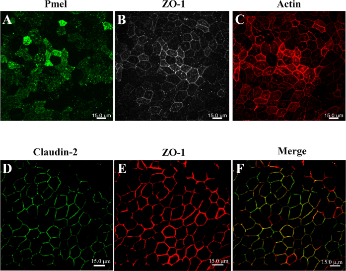
ARPE-19 cells at passages 9 to 12 grown in DMEM containing high glucose and pyruvate with 1% fetal bovine serum were differentiated for up to 4 months. Immunocytochemistry was performed on ARPE-19 cells grown on filters. Total RNA extracted from ARPE-19 cells cultured for either 4 days or 4 months was used for RNA sequencing (RNA-Seq) analysis using a 2 × 50 bp paired end protocol. The RNA-Seq data were analyzed to identify the affected pathways and recognize shared ontological classification among differentially expressed genes. RPE-specific mRNAs and miRNAs were assessed with quantitative real-time (RT)-PCR, and proteins with western blotting.
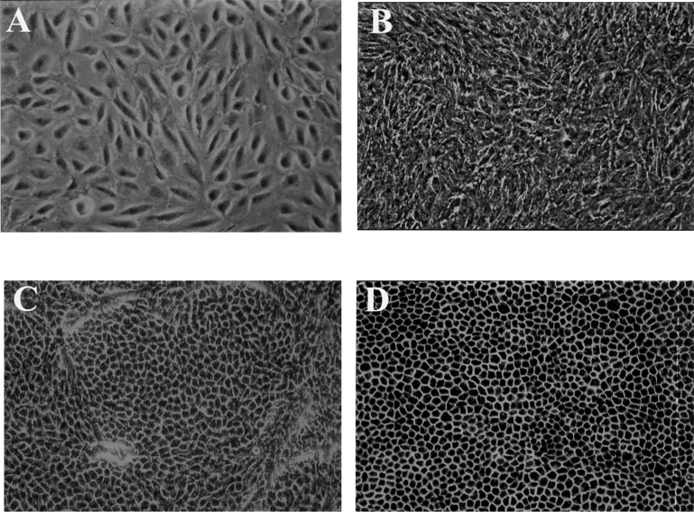
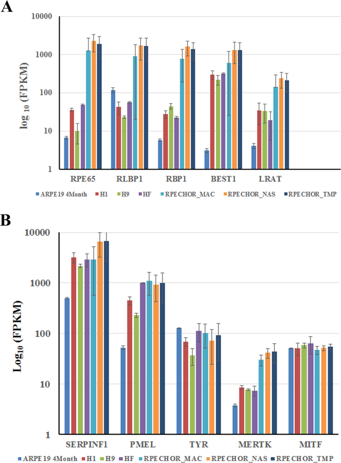
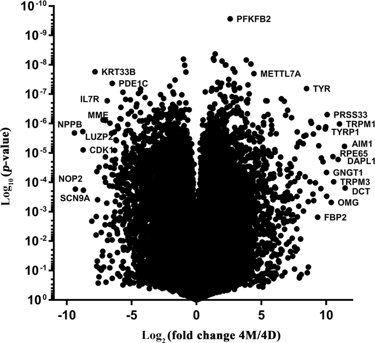
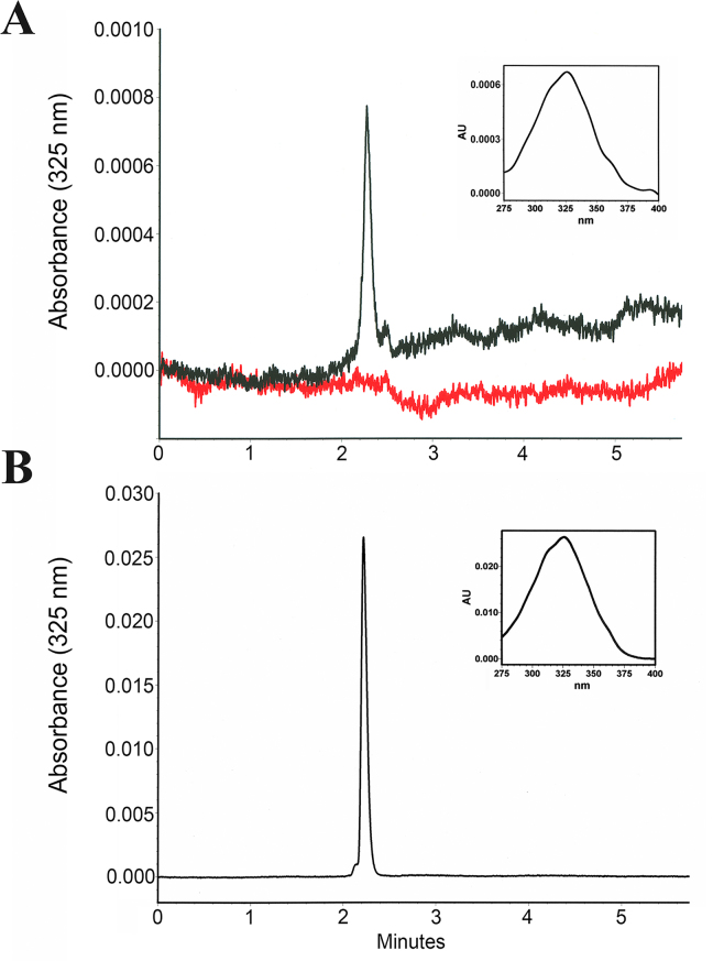
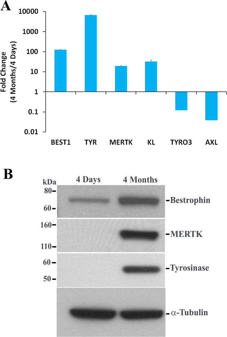
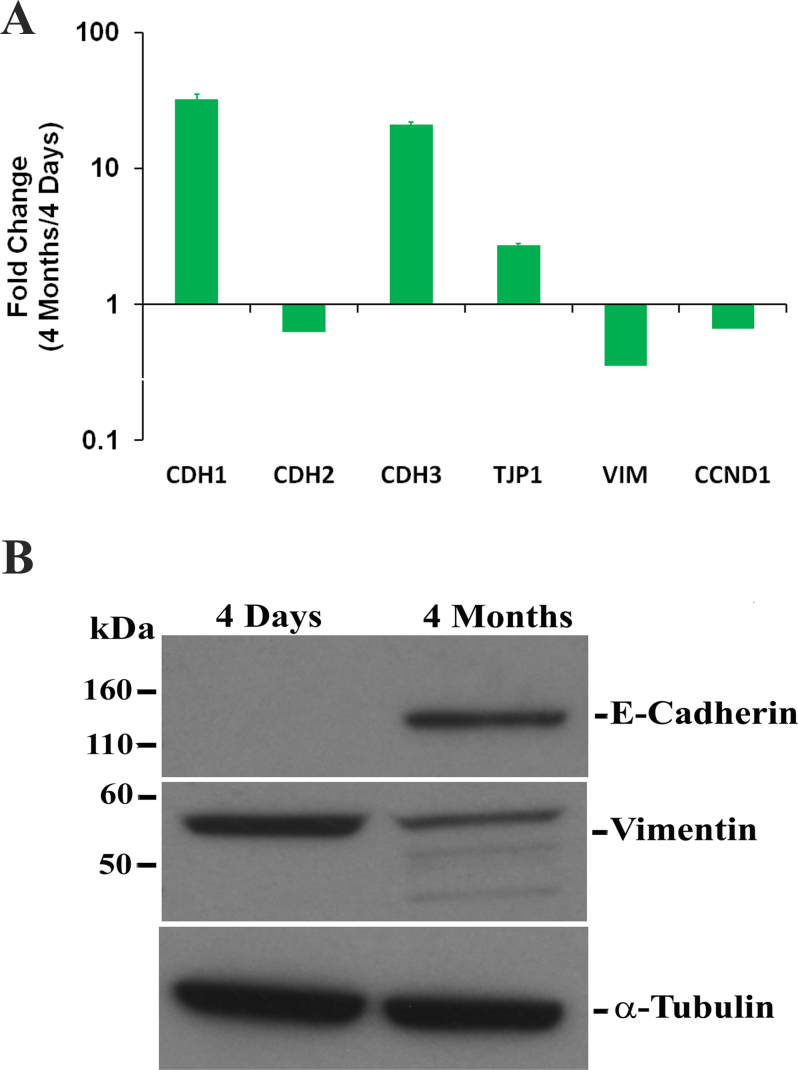
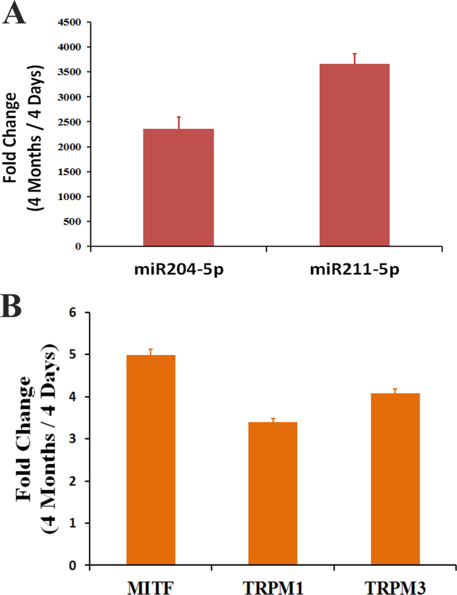
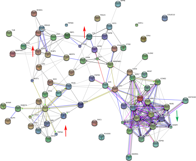
ARPE-19 cells grown for 4 months developed the classic native RPE phenotype with heavy pigmentation. RPE-expressed genes, including , , and , as well as miR-204/211, were greatly increased in the ARPE-19 cells maintained at confluence for 4 months. The RNA-Seq analysis provided a comprehensive view of the relative abundance and differential expression of the genes in the differentiated ARPE-19 cells. Of the 16,757 genes with detectable signals, nearly 1,681 genes were upregulated, and 1,629 genes were downregulated with a fold change of 2.5 or more differences between 4 months and 4 days of culture. Gene Ontology analysis showed that the upregulated genes were associated with visual cycle, phagocytosis, pigment synthesis, cell differentiation, and RPE-related transcription factors. The majority of the downregulated genes play a role in cell cycle and proliferation.
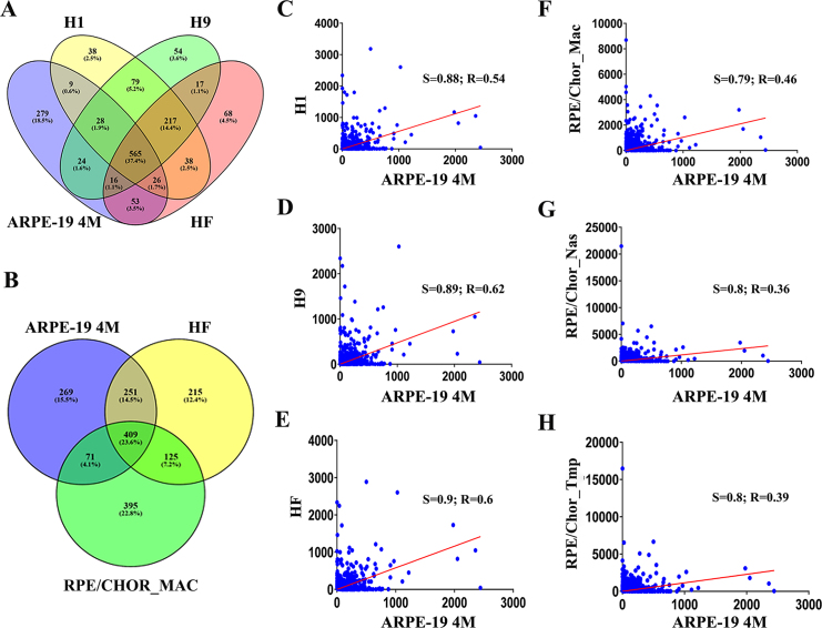
The ARPE-19 cells cultured for 4 months developed a phenotype characteristic of native RPE and expressed proteins, mRNAs, and miRNAs characteristic of the RPE. Comparison of the ARPE-19 RNA-Seq data set with that of primary human fetal RPE, embryonic stem cell-derived RPE, and native RPE revealed an important overall similar expression ratio among all the models and native tissue. However, none of the cultured models reached the absolute values in the native tissue. The results of this study demonstrate that low-passage ARPE-19 cells can express genes specific to native human RPE cells when appropriately cultured and differentiated.
视网膜色素上皮(RPE)细胞系ARPE - 19为天然RPE提供了一种可靠且广泛使用的替代物。然而,由于这些细胞在多次传代后失去其特化表型,复制天然RPE表型变得更加困难。使这个问题更加复杂的是,ARPE - 19细胞以未分化状态被广泛用于尝试模拟RPE功能。我们希望确定合适的培养条件和分化是否能使ARPE - 19恢复与RPE相适应的基因和蛋白质表达,以及类似于天然RPE的功能和形态表型。我们将长期培养的ARPE - 19细胞的转录组与原代及其他人类RPE细胞的转录组进行比较,以评估前者相对于后者的固有可塑性。
在含高糖和丙酮酸钠以及1%胎牛血清的DMEM中培养至第9至12代的ARPE - 19细胞分化长达4个月。对生长在滤膜上的ARPE - 19细胞进行免疫细胞化学分析。从培养4天或4个月的ARPE - 19细胞中提取的总RNA用于使用2×50 bp双端协议的RNA测序(RNA - Seq)分析。对RNA - Seq数据进行分析以识别受影响的途径,并识别差异表达基因之间共享的本体分类。用定量实时(RT)-PCR评估RPE特异性mRNA和miRNA,用蛋白质印迹法评估蛋白质。
培养4个月的ARPE - 19细胞形成了具有大量色素沉着的典型天然RPE表型。在汇合状态下维持4个月的ARPE - 19细胞中,包括 、 和 以及miR - 204/211在内的RPE表达基因大幅增加。RNA - Seq分析全面展示了分化的ARPE - 19细胞中基因的相对丰度和差异表达。在16757个有可检测信号的基因中,培养4个月与培养4天时相比,近1681个基因上调,1629个基因下调,变化倍数为2.5或更大。基因本体分析表明,上调基因与视觉循环、吞噬作用、色素合成、细胞分化以及RPE相关转录因子有关。大多数下调基因在细胞周期和增殖中起作用。
培养4个月的ARPE - 19细胞形成了天然RPE的特征性表型,并表达了RPE特征性的蛋白质、mRNA和miRNA。将ARPE - 19的RNA - Seq数据集与原代人胎儿RPE、胚胎干细胞衍生的RPE和天然RPE的数据集进行比较,发现在所有模型和天然组织之间存在重要的总体相似表达比例。然而,没有一个培养模型达到天然组织中的绝对值。本研究结果表明,低传代的ARPE - 19细胞在适当培养和分化时可以表达天然人类RPE细胞特有的基因。