Modinos G, McLaughlin A, Egerton A, McMullen K, Kumari V, Barker G J, Keysers C, Williams S C R
Institute of Psychiatry, Psychology and Neuroscience, King's College London, London, UK.
Centre for Brain Health, University of British Columbia, Vancouver, BC, Canada.
Transl Psychiatry. 2017 Apr 4;7(4):e1083. doi: 10.1038/tp.2017.53.
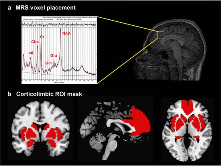
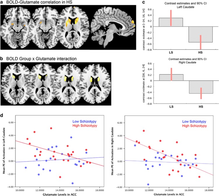
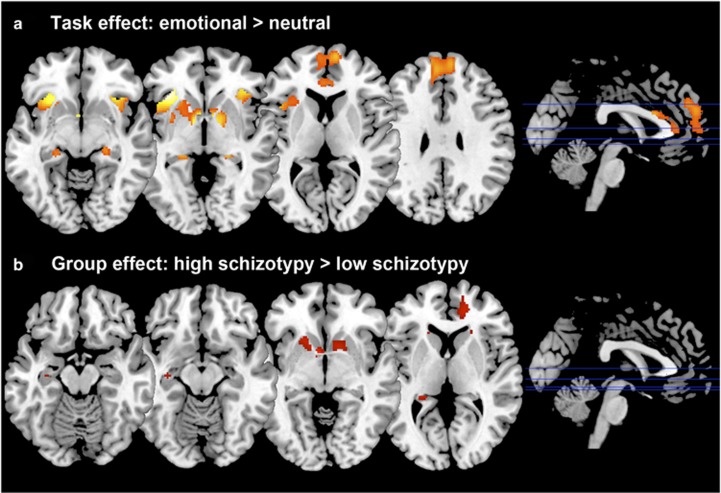
Animal models and human neuroimaging studies suggest that altered levels of glutamatergic metabolites within a corticolimbic circuit have a major role in the pathophysiology of schizophrenia. Rodent models propose that prefrontal glutamate dysfunction could lead to amygdala hyper-response to environmental stress and underlie hippocampal overdrive in schizophrenia. Here we determine whether changes in brain glutamate are present in individuals with high schizotypy (HS), which refers to the presence of schizophrenia-like characteristics in healthy individuals, and whether glutamate levels are related to altered corticolimbic response to emotion. Twenty-one healthy HS subjects and 22 healthy subjects with low schizotypy (LS) were selected based on their Oxford and Liverpool Inventory of Feelings and Experiences rating. Glutamate levels were measured in the anterior cingulate cortex (ACC) using proton magnetic resonance spectroscopy, followed by a functional magnetic resonance imaging (fMRI) scan to measure corticolimbic response during emotional processing. fMRI results and fMRI × glutamate interactions were considered significant after voxel-wise P<0.05 family-wise error correction. While viewing emotional pictures, HS individuals showed greater activation than did subjects with LS in the caudate, and marginally in the ACC, hippocampus, medial prefrontal cortex (MPFC) and putamen. Although no between-group differences were found in glutamate concentrations, within the HS group ACC glutamate was negatively correlated with striatal activation (left: z=4.30, P=0.004 and right: z=4.12 P=0.008 caudate; left putamen: z=3.89, P=0.018) and marginally with MPFC (z=3.55, P=0.052) and amygdala (left: z=2.88, P=0.062; right: z=2.79, P=0.079), correlations that were not present in LS subjects. These findings provide, to our knowledge, the first evidence that brain glutamate levels are associated with hyper-responsivity in brain regions thought to be critical in the pathophysiology of psychosis.
动物模型和人类神经影像学研究表明,皮质边缘回路中谷氨酸能代谢物水平的改变在精神分裂症的病理生理学中起主要作用。啮齿动物模型表明,前额叶谷氨酸功能障碍可能导致杏仁核对环境压力的过度反应,并成为精神分裂症中海马体过度活动的基础。在这里,我们确定高精神分裂症型特质(HS)个体(指健康个体中存在类似精神分裂症的特征)的大脑谷氨酸是否发生变化,以及谷氨酸水平是否与皮质边缘对情绪的反应改变有关。根据牛津和利物浦情感与体验量表评分,选取了21名健康的高精神分裂症型特质受试者和22名低精神分裂症型特质(LS)的健康受试者。使用质子磁共振波谱在前扣带回皮质(ACC)测量谷氨酸水平,随后进行功能磁共振成像(fMRI)扫描,以测量情绪处理过程中的皮质边缘反应。在体素水平P<0.05的家族性错误校正后,fMRI结果和fMRI×谷氨酸相互作用被认为具有显著性。在观看情绪图片时,高精神分裂症型特质个体在尾状核的激活程度高于低精神分裂症型特质受试者,在前扣带回皮质、海马体、内侧前额叶皮质(MPFC)和壳核的激活程度略高。虽然两组之间在谷氨酸浓度上没有差异,但在高精神分裂症型特质组中,前扣带回皮质谷氨酸与纹状体激活呈负相关(左侧:z=4.30,P=0.004;右侧:z=4.12,尾状核P=0.008;左侧壳核:z=3.89,P=0.018),与内侧前额叶皮质呈微弱负相关(z=3.55,P=0.052),与杏仁核呈微弱负相关(左侧:z=2.88,P=0.062;右侧:z=2.79,P=0.079),而在低精神分裂症型特质受试者中不存在这些相关性。据我们所知,这些发现首次证明大脑谷氨酸水平与被认为在精神病病理生理学中至关重要的脑区的高反应性有关。