Stadler Krystina L, Pease Anthony P, Ballegeer Elizabeth A
Department of Small Animal Clinical Sciences, Michigan State University College of Veterinary Medicine , East Lansing, MI , USA.
Front Vet Sci. 2017 Mar 21;4:41. doi: 10.3389/fvets.2017.00041. eCollection 2017.
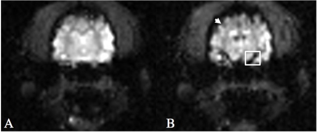
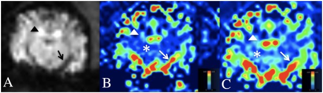
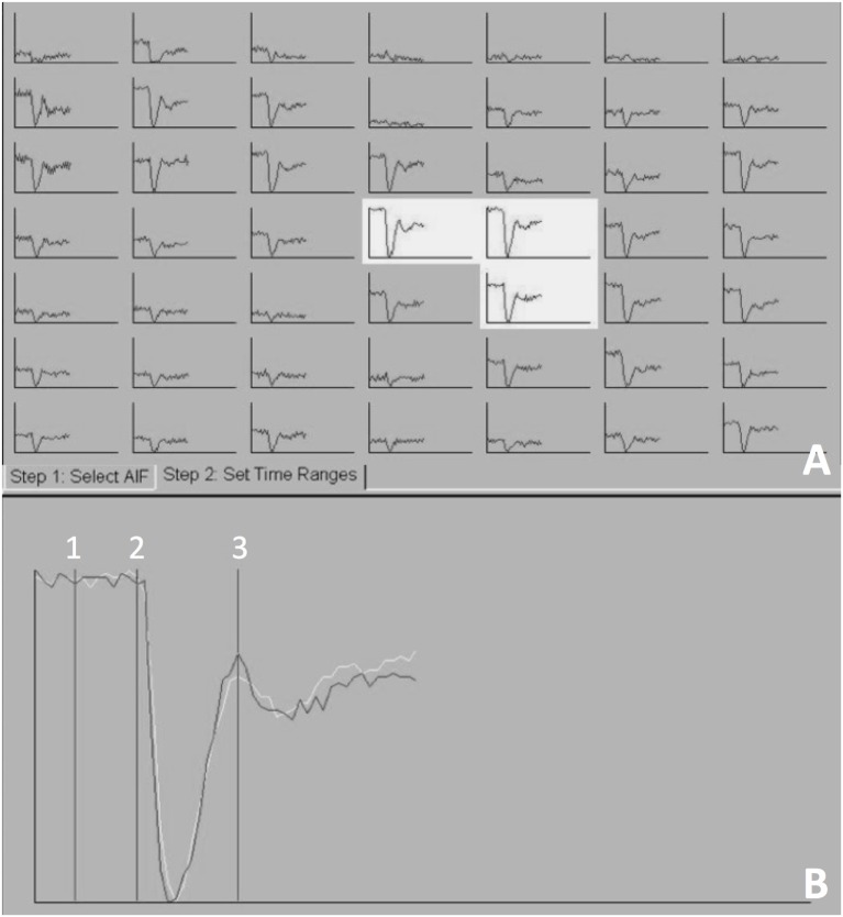
Perfusion magnetic resonance imaging (MRI), specifically dynamic susceptibility MRI (DSC-MRI) is routinely performed as a supplement to conventional MRI in human medicine for patients with intracranial neoplasia and cerebrovascular events. There is minimal data on the use of DSC-MRI in veterinary patients and a DSC-MRI protocol in the veterinary patient has not been described. Sixteen normal dogs, 6 years or older were recruited for this study. The sample population included 11 large dogs (>11 kg) and 5 small dogs (<11 kg). DSC-MRI was performed on a 1.5-T MRI using an adjusted protocol inherent to the MRI. Contrast media was injected using an automatic power injector. Injections were made after five MR measurements were obtained. Following image acquisition, an arterial input function (AIF) graph mapping the transit time of contrast within the cerebral arteries was generated. The manually selected time points along this graph were used to compute perfusion maps. A dose and rate of 0.1 mmol/kg gadolinium-based contrast media at 3 ml/s followed by 10 ml saline flush at 3 ml/s was used in all dogs greater than 11 kg. In all dogs >11 kg, a useable AIF and perfusion map was generated. One dog less than 11 kg received the same contrast dose and rate. In this patient, the protocol did not generate a useable AIF. The remainder of the dogs less than 11 kg followed a protocol of 0.2 mmol/kg gadolinium-based contrast media at 1.5 ml/s with a 10 ml saline flush at 1.5 ml/s. A useable AIF and perfusion map was generated in the remaining dogs <11 kg using the higher contrast dose and slower rate protocol. This study establishes a contrast dose and administration rate for canine DSC-MRI imaging that is different in dogs greater than 11 kg compared to dogs less than 11 kg. These protocols may be used for future applications to evaluate hemodynamic disturbances in canine intracranial pathology.
灌注磁共振成像(MRI),特别是动态磁敏感对比增强MRI(DSC-MRI),在人类医学中常用于颅内肿瘤和脑血管疾病患者,作为传统MRI的补充手段。关于DSC-MRI在兽医患者中的应用数据极少,且尚未描述针对兽医患者的DSC-MRI方案。本研究招募了16只6岁及以上的正常犬。样本群体包括11只大型犬(体重>11千克)和5只小型犬(体重<11千克)。使用MRI固有的调整方案在1.5-T MRI上进行DSC-MRI检查。使用自动注射器注入造影剂。在获得5次MR测量结果后进行注射。图像采集后,生成动脉输入函数(AIF)图,描绘造影剂在脑动脉内的通过时间。沿着该图手动选择的时间点用于计算灌注图。所有体重>11千克的犬使用剂量为0.1 mmol/kg的钆基造影剂,注射速率为3 ml/s,随后以3 ml/s的速率注射10 ml生理盐水冲洗液。所有体重>11千克的犬均生成了可用的AIF和灌注图。1只体重<11千克的犬接受了相同的造影剂剂量和速率。在这只犬中,该方案未生成可用的AIF。其余体重<11千克的犬遵循的方案是使用剂量为0.2 mmol/kg的钆基造影剂,注射速率为1.5 ml/s,随后以1.5 ml/s的速率注射10 ml生理盐水冲洗液。使用更高的造影剂剂量和更慢的注射速率方案,在其余体重<11千克的犬中生成了可用的AIF和灌注图。本研究确定了犬DSC-MRI成像的造影剂剂量和给药速率,体重>11千克的犬与体重<11千克的犬有所不同。这些方案可用于未来评估犬颅内病变血流动力学紊乱的应用中。