Alqhtani N R, Logan N J, Meghji S, Leeson R, Brett P M
University College London, Eastman Dental Institute, 256 Gray's Inn Road, London WC1X 8LD, UK; Department of Oral and Maxillofacial Surgery and Diagnostic Sciences, College of Dentistry, Sattam bin Abdulaziz University, AlKharj, Saudi Arabia.
University College London, Eastman Dental Institute, 256 Gray's Inn Road, London WC1X 8LD, UK.
Bone Rep. 2017 Feb 16;6:64-69. doi: 10.1016/j.bonr.2017.02.002. eCollection 2017 Jun.
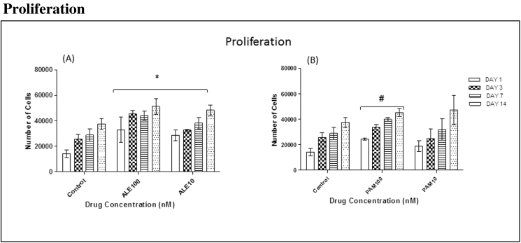
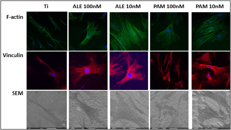
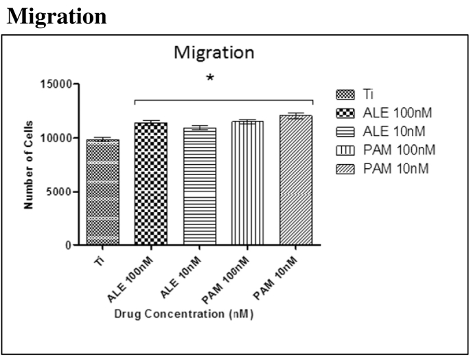
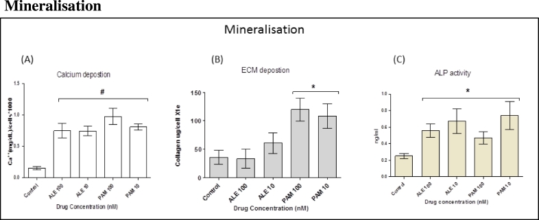
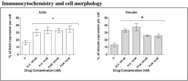
Since the 1980s, titanium (Ti) implants have been routinely used to replace missing teeth. This success is mainly due to the good biocompatibility of Ti and the phenomenon of osseointegration, with very early events at implant placement being important in determining good osseointegration. However, enhancing implant performance with coatings such as hydroxyapatite (HA) and calcium phosphate has proved largely unsuccessful. Human mesenchymal stem cells (hMSCs) are the first osteogenic cells to colonise implant surfaces and offer a target for enhancing osseointegration. We previously reported that small doses of bisphosphonate (BP) may play an integral role in enhancing hMSC proliferation and osteogenic differentiation. The aim of this study is to investigate whether small doses of bisphosphonates enhance proliferation and osteogenic differentiation of hMSCs on Ti surfaces, to enhance bone osseointegration and to accelerate wound healing around the implant surface. Our data suggests that treating cells with small doses of BP (100 nM & 10 nM) induces significant hMSC stimulation of osteogenic markers including calcium, collagen type I and ALP compared to control group on titanium surfaces (P < 0.05). In addition, cell proliferation and migration were significantly enhanced on titanium surfaces (P < 0.05).
自20世纪80年代以来,钛(Ti)植入物一直被常规用于替代缺失的牙齿。这一成功主要归因于钛良好的生物相容性以及骨整合现象,植入时的早期事件对于确定良好的骨整合非常重要。然而,事实证明,用羟基磷灰石(HA)和磷酸钙等涂层来提高植入物性能在很大程度上并不成功。人间充质干细胞(hMSCs)是最早在植入物表面定植的成骨细胞,为增强骨整合提供了一个靶点。我们之前报道过,小剂量双膦酸盐(BP)可能在增强hMSC增殖和成骨分化中发挥不可或缺的作用。本研究的目的是调查小剂量双膦酸盐是否能增强hMSCs在钛表面的增殖和成骨分化,以增强骨整合并加速植入物表面周围的伤口愈合。我们的数据表明,与钛表面的对照组相比,用小剂量BP(100 nM和10 nM)处理细胞可显著刺激hMSC产生包括钙、I型胶原蛋白和碱性磷酸酶在内的成骨标志物(P < 0.05)。此外,钛表面的细胞增殖和迁移也显著增强(P < 0.05)。