• 文献检索
  • 文档翻译
  • 深度研究
  • 学术资讯
  • Suppr Zotero 插件Zotero 插件
  • 邀请有礼
  • 套餐&价格
  • 历史记录
应用&插件
Suppr Zotero 插件Zotero 插件浏览器插件Mac 客户端Windows 客户端微信小程序
定价
高级版会员购买积分包购买API积分包
服务
文献检索文档翻译深度研究API 文档MCP 服务
关于我们
关于 Suppr公司介绍联系我们用户协议隐私条款
关注我们

Suppr 超能文献

核心技术专利:CN118964589B侵权必究
粤ICP备2023148730 号-1Suppr @ 2026

文献检索

告别复杂PubMed语法,用中文像聊天一样搜索,搜遍4000万医学文献。AI智能推荐,让科研检索更轻松。

立即免费搜索

文件翻译

保留排版,准确专业,支持PDF/Word/PPT等文件格式,支持 12+语言互译。

免费翻译文档

深度研究

AI帮你快速写综述,25分钟生成高质量综述,智能提取关键信息,辅助科研写作。

立即免费体验

使用表观扩散系数(ADC)参数反应图鉴别胶质母细胞瘤中的假性进展和真性进展

Differentiation of pseudoprogression and real progression in glioblastoma using ADC parametric response maps.

作者信息

Reimer Caroline, Deike Katerina, Graf Markus, Reimer Peter, Wiestler Benedikt, Floca Ralf Omar, Kickingereder Philipp, Schlemmer Heinz-Peter, Wick Wolfgang, Bendszus Martin, Radbruch Alexander

机构信息

Department of Neuroradiology, University of Heidelberg Medical Center, Heidelberg, Germany.

Department of Radiology, Deutsches Krebsforschungszentrum (DKFZ), Heidelberg, Germany.

出版信息

PLoS One. 2017 Apr 6;12(4):e0174620. doi: 10.1371/journal.pone.0174620. eCollection 2017.

DOI:10.1371/journal.pone.0174620
PMID:28384170
原文链接:https://pmc.ncbi.nlm.nih.gov/articles/PMC5383222/
Abstract

PURPOSE

The purpose of this study was to investigate whether a voxel-wise analysis of apparent diffusion coefficient (ADC) values may differentiate between progressive disease (PD) and pseudoprogression (PsP) in patients with high-grade glioma using the parametric response map, a newly introduced postprocessing tool.

METHODS

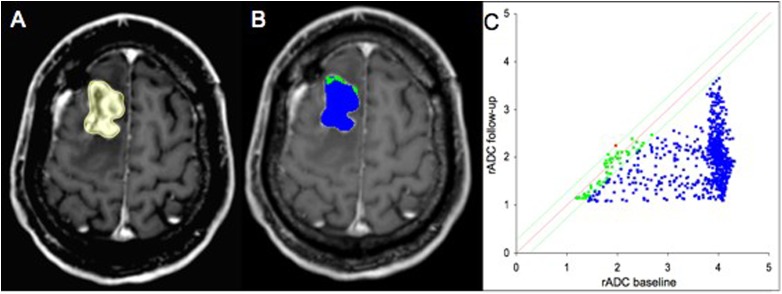
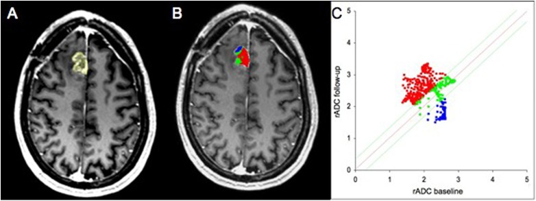
Twenty-eight patients with proven PD and seven patients with PsP were identified in this retrospective feasibility study. For all patients ADC baseline and follow-up maps on four subsequent MRIs were available. ADC maps were coregistered on contrast enhanced T1-weighted follow-up images. Subsequently, enhancement in the follow-up contrast enhanced T1-weighted image was manually delineated and a reference region of interest (ROI) was drawn in the contralateral white matter. Both ROIs were transferred to the ADC images. Relative ADC (rADC) (baseline)/reference ROI values and rADC (follow up)/reference ROI values were calculated for each voxel within the ROI. The corresponding voxels of rADC (follow up) and rADC (baseline) were subtracted and the percentage of all voxels within the ROI that exceeded the threshold of 0.25 was quantified.

RESULTS

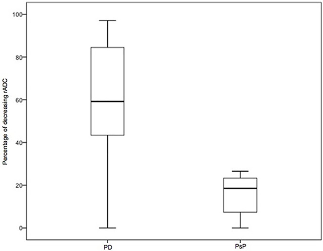
rADC voxels showed a decrease of 59.2% (1st quartile (Q1) 36.7; 3rd quartile (Q3) 78.6) above 0.25 in patients with PD and 18.6% (Q1 3.04; Q3 26.5) in patients with PsP (p = 0.005). Receiver operating characteristic curve analysis showed the optimal decreasing rADC cut-off value for identifying PD of > 27.05% (area under the curve 0.844±0.065, sensitivity 0.86, specificity 0.86, p = 0.014).

CONCLUSION

This feasibility study shows that the assessment of rADC using parametric response maps might be a promising approach to contribute to the differentiation between PD and PsP. Further research in larger patient cohorts is necessary to finally determine its clinical utility.

摘要

目的

本研究旨在探讨使用一种新引入的后处理工具——参数反应映射,对高级别胶质瘤患者表观扩散系数(ADC)值进行体素分析,是否能够区分疾病进展(PD)和假性进展(PsP)。

方法

在这项回顾性可行性研究中,确定了28例经证实为PD的患者和7例PsP患者。所有患者均有后续4次MRI检查的ADC基线图和随访图。将ADC图与增强T1加权随访图像进行配准。随后,手动勾勒出随访增强T1加权图像中的强化区域,并在对侧白质中绘制一个感兴趣参考区域(ROI)。将两个ROI都转移到ADC图像上。计算ROI内每个体素的相对ADC(rADC)(基线)/参考ROI值和rADC(随访)/参考ROI值。将rADC(随访)和rADC(基线)的相应体素相减,并对ROI内超过0.25阈值的所有体素的百分比进行量化。

结果

PD患者中,rADC体素在0.25以上下降了59.2%(第一四分位数(Q1)36.7;第三四分位数(Q3)78.6),PsP患者中下降了18.6%(Q1 3.04;Q3 26.5)(p = 0.005)。受试者操作特征曲线分析显示,识别PD的最佳rADC下降临界值>27.05%(曲线下面积0.844±0.065,敏感性0.86,特异性0.86,p = 0.014)。

结论

这项可行性研究表明,使用参数反应映射评估rADC可能是一种有助于区分PD和PsP的有前景方法。需要在更大患者队列中进行进一步研究,以最终确定其临床实用性。

https://cdn.ncbi.nlm.nih.gov/pmc/blobs/1482/5383222/4b73a4a1ae60/pone.0174620.g003.jpg
https://cdn.ncbi.nlm.nih.gov/pmc/blobs/1482/5383222/3ef4d85f68fb/pone.0174620.g001.jpg
https://cdn.ncbi.nlm.nih.gov/pmc/blobs/1482/5383222/7c6d78aebf4a/pone.0174620.g002.jpg
https://cdn.ncbi.nlm.nih.gov/pmc/blobs/1482/5383222/4b73a4a1ae60/pone.0174620.g003.jpg
https://cdn.ncbi.nlm.nih.gov/pmc/blobs/1482/5383222/3ef4d85f68fb/pone.0174620.g001.jpg
https://cdn.ncbi.nlm.nih.gov/pmc/blobs/1482/5383222/7c6d78aebf4a/pone.0174620.g002.jpg
https://cdn.ncbi.nlm.nih.gov/pmc/blobs/1482/5383222/4b73a4a1ae60/pone.0174620.g003.jpg

相似文献

1
Differentiation of pseudoprogression and real progression in glioblastoma using ADC parametric response maps.使用表观扩散系数(ADC)参数反应图鉴别胶质母细胞瘤中的假性进展和真性进展
PLoS One. 2017 Apr 6;12(4):e0174620. doi: 10.1371/journal.pone.0174620. eCollection 2017.
2
Differentiation of true progression from pseudoprogression in glioblastoma treated with radiation therapy and concomitant temozolomide: comparison study of standard and high-b-value diffusion-weighted imaging.在接受放疗和替莫唑胺联合治疗的胶质母细胞瘤中,从假性进展中区分真性进展:标准和高 b 值扩散加权成像的对比研究。
Radiology. 2013 Dec;269(3):831-40. doi: 10.1148/radiol.13122024. Epub 2013 Oct 28.
3
Comparison of two region-of-interest placement methods for histogram analysis of apparent diffusion coefficient maps for glioma grading.两种感兴趣区放置方法在胶质瘤分级表观扩散系数图直方图分析中的比较。
Clin Ter. 2024 May-Jun;175(3):128-136. doi: 10.7417/CT.2024.5053.
4
True progression versus pseudoprogression in the treatment of glioblastomas: a comparison study of normalized cerebral blood volume and apparent diffusion coefficient by histogram analysis.治疗胶质母细胞瘤中的真性进展与假性进展:通过直方图分析比较归一化脑血容量和表观扩散系数。
Korean J Radiol. 2013 Jul-Aug;14(4):662-72. doi: 10.3348/kjr.2013.14.4.662. Epub 2013 Jul 17.
5
Lymphomas and glioblastomas: differences in the apparent diffusion coefficient evaluated with high b-value diffusion-weighted magnetic resonance imaging at 3T.淋巴瘤和胶质母细胞瘤:3T 高 b 值扩散加权磁共振成像评估表观扩散系数的差异。
Eur J Radiol. 2012 Feb;81(2):339-44. doi: 10.1016/j.ejrad.2010.11.005. Epub 2010 Dec 3.
6
Relative apparent diffusion coefficient: a promising tool to differentiate metastatic from benign lymph nodes in animal models.相对表观扩散系数:一种有前途的工具,可用于区分动物模型中的转移性和良性淋巴结。
Chin Med J (Engl). 2011 Sep;124(18):2907-10.
7
Prognostic value of combined visualization of MR diffusion and perfusion maps in glioblastoma.磁共振扩散与灌注成像联合可视化在胶质母细胞瘤中的预后价值
J Neurooncol. 2016 Feb;126(3):463-72. doi: 10.1007/s11060-015-1982-z. Epub 2015 Oct 30.
8
Glioma grading using apparent diffusion coefficient map: application of histogram analysis based on automatic segmentation.基于自动分割的直方图分析在利用表观扩散系数图进行胶质瘤分级中的应用
NMR Biomed. 2014 Sep;27(9):1046-52. doi: 10.1002/nbm.3153. Epub 2014 Jul 7.
9
Cerebral schistosomiasis: diffusion-weighted imaging helps to differentiate from brain glioma and metastasis.脑血吸虫病:弥散加权成像有助于与脑胶质瘤和转移瘤相鉴别。
Acta Radiol. 2017 Nov;58(11):1371-1377. doi: 10.1177/0284185116687173. Epub 2017 Jan 16.
10
Assessment of histogram analysis in distinguishing between low-grade and high-grade brainstem glioma.评估直方图分析在低级别和高级别脑干胶质瘤鉴别中的作用。
Clin Ter. 2024 Sep-Oct;175(5):296-306. doi: 10.7417/CT.2024.5134.

引用本文的文献

1
Association between the Anatomical Location of Glioblastoma and Its Evaluation with Clinical Considerations: A Systematic Review and Meta-Analysis.胶质母细胞瘤的解剖位置与其临床评估之间的关联:一项系统评价和荟萃分析
J Clin Med. 2024 Jun 13;13(12):3460. doi: 10.3390/jcm13123460.
2
Validation of daily 0.35 T diffusion-weighted MRI for MRI-guided glioblastoma radiotherapy.每日 0.35T 弥散加权 MRI 用于 MRI 引导下脑胶质瘤放疗的验证。
Med Phys. 2024 Aug;51(8):5386-5398. doi: 10.1002/mp.17067. Epub 2024 Apr 8.
3
Undetected pseudoprogressions in the CeTeG/NOA-09 trial: hints from postprogression survival and MRI analyses.

本文引用的文献

1
MR Perfusion-derived Hemodynamic Parametric Response Mapping of Bevacizumab Efficacy in Recurrent Glioblastoma.基于灌注 MRI 的贝伐珠单抗治疗复发性胶质母细胞瘤的血流动力学参数反应映射。
Radiology. 2016 May;279(2):542-52. doi: 10.1148/radiol.2015151172. Epub 2015 Nov 16.
2
Prognostic value of combined visualization of MR diffusion and perfusion maps in glioblastoma.磁共振扩散与灌注成像联合可视化在胶质母细胞瘤中的预后价值
J Neurooncol. 2016 Feb;126(3):463-72. doi: 10.1007/s11060-015-1982-z. Epub 2015 Oct 30.
3
Differentiating Tumor Progression from Pseudoprogression in Patients with Glioblastomas Using Diffusion Tensor Imaging and Dynamic Susceptibility Contrast MRI.
CeTeG/NOA-09 试验中的未检测到的假性进展:来自进展后生存和 MRI 分析的提示。
J Neurooncol. 2023 Sep;164(3):607-616. doi: 10.1007/s11060-023-04444-x. Epub 2023 Sep 20.
4
Differentiating tumour progression from pseudoprogression in glioblastoma patients: a monoexponential, biexponential, and stretched-exponential model-based DWI study.基于单指数、双指数和拉伸指数模型的 DWI 研究:区分胶质母细胞瘤患者的肿瘤进展与假性进展。
BMC Med Imaging. 2023 Sep 11;23(1):119. doi: 10.1186/s12880-023-01082-7.
5
Whole body FDG PET/MR for progression free and overall survival prediction in patients with relapsed/refractory large B-cell lymphomas undergoing CAR T-cell therapy.全身 FDG PET/MR 对接受 CAR T 细胞治疗的复发/难治性大 B 细胞淋巴瘤患者进行无进展生存和总生存预测。
Cancer Imaging. 2022 Dec 27;22(1):76. doi: 10.1186/s40644-022-00513-y.
6
Assessment of the hypervascularized fraction of glioblastomas using a volume analysis of dynamic susceptibility contrast-enhanced MRI may help to identify pseudoprogression.使用动态磁敏感对比增强 MRI 的容积分析评估胶质母细胞瘤的富血管化部分,可能有助于识别假性进展。
PLoS One. 2022 Oct 13;17(10):e0270216. doi: 10.1371/journal.pone.0270216. eCollection 2022.
7
Diffusion imaging could aid to differentiate between glioma progression and treatment-related abnormalities: a meta-analysis.扩散成像有助于鉴别胶质瘤进展与治疗相关异常:一项荟萃分析。
Insights Imaging. 2022 Oct 4;13(1):158. doi: 10.1186/s13244-022-01295-4.
8
Tumor Progression and Treatment-Related Changes: Radiological Diagnosis Challenges for the Evaluation of Post Treated Glioma.肿瘤进展及与治疗相关的变化:治疗后胶质瘤评估的放射学诊断挑战
Cancers (Basel). 2022 Aug 3;14(15):3771. doi: 10.3390/cancers14153771.
9
Discriminators of pseudoprogression and true progression in high-grade gliomas: A systematic review and meta-analysis.高级别胶质瘤中假性进展和真性进展的鉴别因素:系统评价和荟萃分析。
Sci Rep. 2022 Aug 2;12(1):13258. doi: 10.1038/s41598-022-16726-x.
10
High-Grade Glioma Treatment Response Monitoring Biomarkers: A Position Statement on the Evidence Supporting the Use of Advanced MRI Techniques in the Clinic, and the Latest Bench-to-Bedside Developments. Part 1: Perfusion and Diffusion Techniques.高级别胶质瘤治疗反应监测生物标志物:关于支持在临床中使用先进MRI技术的证据以及最新的从 bench 到 bedside 进展的立场声明。第1部分:灌注和扩散技术。
Front Oncol. 2022 Mar 3;12:810263. doi: 10.3389/fonc.2022.810263. eCollection 2022.
利用扩散张量成像和动态磁敏感对比磁共振成像鉴别胶质母细胞瘤患者的肿瘤进展与假性进展
AJNR Am J Neuroradiol. 2016 Jan;37(1):28-36. doi: 10.3174/ajnr.A4474. Epub 2015 Oct 8.
4
Diagnosis of pseudoprogression in patients with glioblastoma using O-(2-[18F]fluoroethyl)-L-tyrosine PET.使用O-(2-[¹⁸F]氟乙基)-L-酪氨酸PET对胶质母细胞瘤患者假性进展的诊断
Eur J Nucl Med Mol Imaging. 2015 Apr;42(5):685-95. doi: 10.1007/s00259-014-2959-4. Epub 2014 Nov 20.
5
Glioblastoma treated with concurrent radiation therapy and temozolomide chemotherapy: differentiation of true progression from pseudoprogression with quantitative dynamic contrast-enhanced MR imaging.胶质母细胞瘤行同期放化疗治疗:定量动态对比增强磁共振成像鉴别真性进展与假性进展。
Radiology. 2015 Mar;274(3):830-40. doi: 10.1148/radiol.14132632. Epub 2014 Oct 21.
6
Pseudoprogression in patients with glioblastoma: clinical relevance despite low incidence.胶质母细胞瘤患者的假性进展:尽管发病率低但具有临床相关性。
Neuro Oncol. 2015 Jan;17(1):151-9. doi: 10.1093/neuonc/nou129. Epub 2014 Jul 18.
7
Diffusion-Weighted MRI as a Biomarker of Tumor Radiation Treatment Response Heterogeneity: A Comparative Study of Whole-Volume Histogram Analysis versus Voxel-Based Functional Diffusion Map Analysis.扩散加权 MRI 作为肿瘤放射治疗反应异质性的生物标志物:全容积直方图分析与体素基功能扩散图分析的比较研究。
Transl Oncol. 2013 Oct 1;6(5):554-61. doi: 10.1593/tlo.13532. eCollection 2013.
8
Prediction of pseudoprogression in patients with glioblastomas using the initial and final area under the curves ratio derived from dynamic contrast-enhanced T1-weighted perfusion MR imaging.利用动态对比增强T1加权灌注磁共振成像得出的曲线下初始面积与最终面积之比预测胶质母细胞瘤患者的假性进展
AJNR Am J Neuroradiol. 2013 Dec;34(12):2278-86. doi: 10.3174/ajnr.A3634. Epub 2013 Jul 4.
9
Discriminating radiation necrosis from tumor progression in gliomas: a systematic review what is the best imaging modality?鉴别脑胶质瘤中的放射性坏死与肿瘤进展:哪种影像学方法最佳?系统综述
J Neurooncol. 2013 Apr;112(2):141-52. doi: 10.1007/s11060-013-1059-9. Epub 2013 Jan 24.
10
Differentiating tumor recurrence from treatment necrosis: a review of neuro-oncologic imaging strategies.肿瘤复发与治疗坏死的鉴别:神经肿瘤影像学策略综述。
Neuro Oncol. 2013 May;15(5):515-34. doi: 10.1093/neuonc/nos307. Epub 2013 Jan 16.