Ehlerding Emily B, England Christopher G, Majewski Rebecca L, Valdovinos Hector F, Jiang Dawei, Liu Glenn, McNeel Douglas G, Nickles Robert J, Cai Weibo
Department of Medical Physics, University of Wisconsin-Madison , Madison, Wisconsin 53705, United States.
Department of Biomedical Engineering, University of Wisconsin-Madison , Madison, Wisconsin 53705, United States.
Mol Pharm. 2017 May 1;14(5):1782-1789. doi: 10.1021/acs.molpharmaceut.7b00056. Epub 2017 Apr 12.
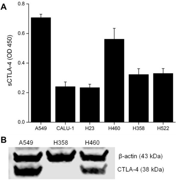
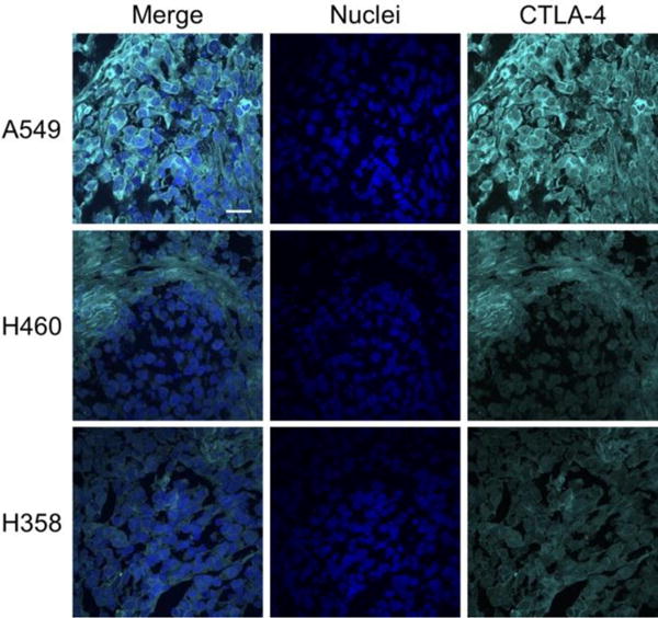
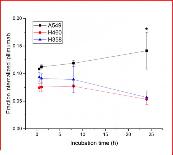
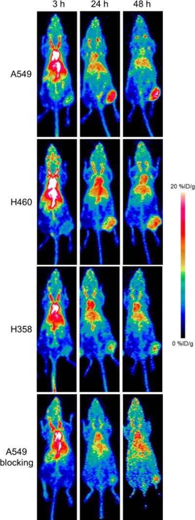
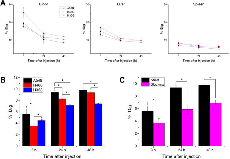
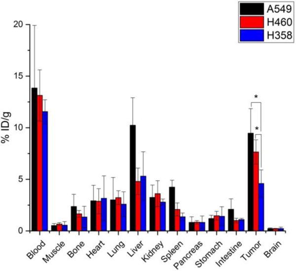
Cytotoxic T-lymphocyte-associated protein 4 (CTLA-4) is expressed on the surface of activated T cells and some tumor cells, and is the target of the clinically approved monoclonal antibody ipilimumab. In this study, we investigate specific binding of radiolabeled ipilimumab to CTLA-4 expressed by human non-small cell lung cancer cells in vivo using positron emission tomography (PET). Ipilimumab was radiolabeled with Cu (t = 12.7 h) through the use of the chelator 1,4,7,10-tetraazacyclododecane-1,4,7,10-tetraacetic acid (DOTA) to formulate Cu-DOTA-ipilimumab. CTLA-4 expression in three non-small cell lung cancer (NSCLC) cell lines (A549, H460, and H358) was verified and quantified by Western blot and enzyme-linked immunosorbent assays (ELISA). A receptor binding assay was utilized to monitor the binding and internalization of Cu-DOTA-ipilimumab in the NSCLC cell lines. Next, the biodistribution of Cu-DOTA-ipilimumab was mapped by longitudinal PET imaging up to 48 h after injection. Ex vivo biodistribution and histological studies were employed to verify PET results. By in vitro analysis, CTLA-4 was found to be expressed on all three NSCLC cell lines with A549 and H358 showing the highest and lowest level of expression, respectively. PET imaging and quantification verified these findings as the tracer accumulated highest in the A549 tumor model (9.80 ± 0.22%ID/g at 48 h after injection; n = 4), followed by H460 and H358 tumors with uptakes of 9.37 ± 0.26%ID/g and 7.43 ± 0.05%ID/g, respectively (n = 4). The specificity of the tracer was verified by injecting excess ipilimumab in A549 tumor-bearing mice, which decreased tracer uptake to 6.90 ± 0.51%ID/g at 48 after injection (n = 4). Ex vivo analysis following the last imaging session also corroborated these findings. Cu-DOTA-ipilimumab showed enhanced and persistent accumulation in CTLA-4-expressing tissues, which will enable researchers further insight into CTLA-4 targeted therapies in the future.
细胞毒性T淋巴细胞相关蛋白4(CTLA-4)表达于活化T细胞和某些肿瘤细胞表面,是临床批准的单克隆抗体伊匹单抗的作用靶点。在本研究中,我们使用正电子发射断层扫描(PET)研究放射性标记的伊匹单抗在体内与人非小细胞肺癌细胞表达的CTLA-4的特异性结合。通过使用螯合剂1,4,7,10-四氮杂环十二烷-1,4,7,10-四乙酸(DOTA)用铜(半衰期 = 12.7小时)对伊匹单抗进行放射性标记,以制备铜- DOTA-伊匹单抗。通过蛋白质印迹法和酶联免疫吸附测定(ELISA)对三种非小细胞肺癌(NSCLC)细胞系(A549、H460和H358)中的CTLA-4表达进行验证和定量。利用受体结合试验监测铜- DOTA-伊匹单抗在NSCLC细胞系中的结合和内化。接下来,通过注射后长达48小时的纵向PET成像绘制铜- DOTA-伊匹单抗的生物分布。采用离体生物分布和组织学研究来验证PET结果。通过体外分析,发现所有三种NSCLC细胞系均表达CTLA-4,其中A549和H358分别显示出最高和最低表达水平。PET成像和定量分析证实了这些发现,因为示踪剂在A549肿瘤模型中积累最高(注射后48小时为9.80±0.22%ID/g;n = 4),其次是H460和H358肿瘤,摄取量分别为9.37±0.26%ID/g和7.43±0.05%ID/g(n = 4)。通过向荷A549肿瘤小鼠注射过量伊匹单抗来验证示踪剂的特异性,这使得注射后48小时示踪剂摄取量降至6.90±0.51%ID/g(n = 4)。最后一次成像后的离体分析也证实了这些发现。铜- DOTA-伊匹单抗在表达CTLA-4的组织中显示出增强且持续的积累,这将使研究人员在未来能够进一步深入了解CTLA-4靶向治疗。