Guangdong Key Laboratory for Biomedical Measurements and Ultrasound Imaging, School of Biomedical Engineering, Health Science Center, Shenzhen University , Shenzhen 518060, China.
University of Wisconsin Carbone Cancer Center , Madison, Wisconsin 53705, United States.
Mol Pharm. 2017 Oct 2;14(10):3239-3247. doi: 10.1021/acs.molpharmaceut.7b00216. Epub 2017 Sep 6.
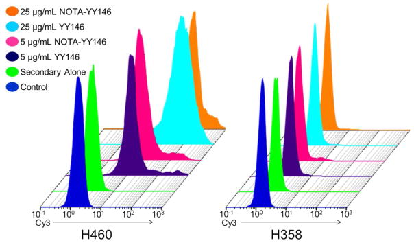
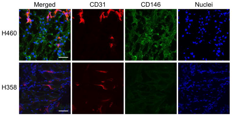
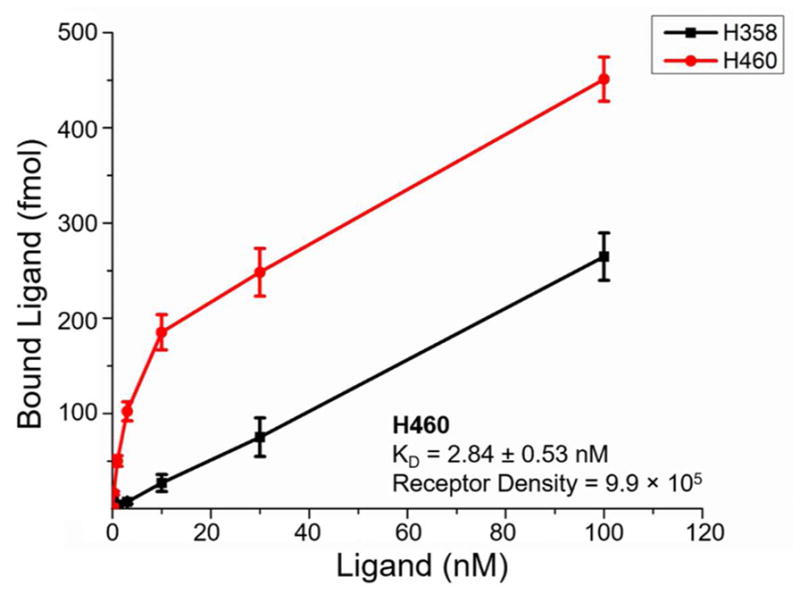
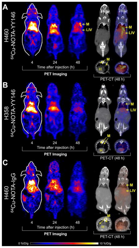
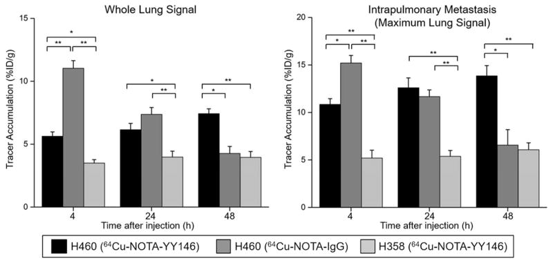
CD146 has been identified as an excellent biomarker for lung cancer as its overexpression in solid tumors has been linked to disease progression, invasion, and metastasis. Previously, our group described a positive correlation between Cu-labeled YY146 uptake and increased expression of CD146 in six human lung cancer cell lines using subcutaneous tumor models. In this study, we investigate a monoclonal antibody called YY146 for immunoPET imaging of CD146 in two intrapulmonary metastasis models of non-small cell lung cancer (NSCLC). The binding and immunoreactivity of the tracer were assessed by in vitro assays. Radiolabeling of YY146 with positron emitting Cu-64 (Cu-NOTA-YY146) enabled PET imaging of intrapulmonary metastasis. Mice were intravenously injected with two million tumor cells, and CT imaging was used to verify the presence of lung metastases. Cu-NOTA-YY146 was injected into tumor-bearing mice, and animals were subjected to PET/CT imaging at 4, 24, and 48 h postinjection. Both the average and maximum lung PET signal intensities were quantified and compared between high and low CD146-expressing metastases. Further validation was accomplished through immunofluorescence imaging of resected tissues with CD31 and CD146. In flow cytometry, YY146 revealed strong binding to CD146 in H460 cells due to its high expression with minimal binding to CD146-low expressing H358 cells. Both YY146 and NOTA-YY146 showed similar binding, suggesting that NOTA conjugation did not elicit any negative effects on its binding affinity. Imaging of Cu-NOTA-YY146 in H460 tumor-bearing mice revealed rapid, persistent, and highly specific tracer accumulation. Uptake of Cu-NOTA-YY146 in the whole lung was calculated for H460 and H358 as 7.43 ± 0.38 and 3.95 ± 0.47% ID/g at 48 h postinjection (n = 4, p < 0.05), and the maximum lung signals were determined to be 13.85 ± 1.07 (H460) and 6.08 ± 0.73% ID/g (H358) at equivalent time points (n = 4, p < 0.05). To ensure the specificity of the tracer, a nonspecific antibody was injected into H460 tumor-bearing mice. Ex vivo biodistribution and immunofluorescence imaging validated the PET findings. In summary, Cu-NOTA-YY146 allowed for successful imaging of CD146-expressing intrapulmonary metastases of NSCLC in mice. This preliminary study provides evidence supporting the future clinical utilization of Cu-NOTA-YY146 for possible treatment monitoring of CD146-targeted therapy or improving patient stratification.
CD146 已被确定为肺癌的优秀生物标志物,因为其在实体瘤中的过度表达与疾病进展、侵袭和转移有关。之前,我们的研究小组在使用皮下肿瘤模型的六种人肺癌细胞系中描述了 Cu 标记的 YY146 摄取与 CD146 表达增加之间的正相关性。在这项研究中,我们研究了一种名为 YY146 的单克隆抗体,用于两种非小细胞肺癌(NSCLC)肺内转移模型中 CD146 的免疫 PET 成像。通过体外测定评估示踪剂的结合和免疫反应性。用正电子发射铜-64(Cu-NOTA-YY146)对 YY146 进行放射性标记,使肺内转移的 PET 成像成为可能。将 200 万个肿瘤细胞静脉注射到小鼠体内,并使用 CT 成像验证肺转移的存在。将 Cu-NOTA-YY146 注射到荷瘤小鼠体内,在注射后 4、24 和 48 小时进行 PET/CT 成像。在高和低 CD146 表达转移之间定量和比较了平均和最大肺 PET 信号强度。通过对切除组织进行 CD31 和 CD146 的免疫荧光成像进一步验证。在流式细胞术中,由于其高表达,YY146 与 H460 细胞中的 CD146 有很强的结合,与 CD146 低表达的 H358 细胞结合很少。YY146 和 NOTA-YY146 均表现出相似的结合,表明 NOTA 缀合没有对其结合亲和力产生任何负面影响。在 H460 荷瘤小鼠中对 Cu-NOTA-YY146 的成像显示出快速、持续和高度特异性示踪剂积累。在注射后 48 小时,对 H460 和 H358 计算的整个肺中的 Cu-NOTA-YY146 摄取量分别为 7.43 ± 0.38 和 3.95 ± 0.47% ID/g(n = 4,p < 0.05),并且在等效时间点确定的最大肺信号分别为 13.85 ± 1.07(H460)和 6.08 ± 0.73% ID/g(H358)(n = 4,p < 0.05)。为了确保示踪剂的特异性,将非特异性抗体注射到 H460 荷瘤小鼠中。离体生物分布和免疫荧光成像验证了 PET 结果。总之,Cu-NOTA-YY146 允许成功地对 NSCLC 小鼠肺内转移的 CD146 表达进行成像。这项初步研究为未来临床应用 Cu-NOTA-YY146 用于 CD146 靶向治疗的可能治疗监测或改善患者分层提供了证据支持。