Nie Jing, Huang Guang-Long, Deng Sheng-Ze, Bao Yun, Liu Ya-Wei, Feng Zhan-Peng, Wang Chao-Hu, Chen Ming, Qi Song-Tao, Pan Jun
Department of NeurosurgeryNanfang Hospital, Southern Medical University, Guangzhou, China.
Nanfang Neurosurgery Research InstitutionNanfang hospital, Southern Medical University, Guangzhou, China.
Endocr Relat Cancer. 2017 Jun;24(6):287-296. doi: 10.1530/ERC-16-0338. Epub 2017 Apr 7.
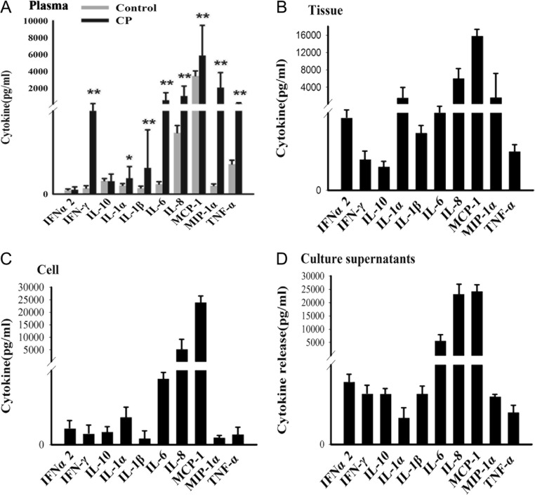
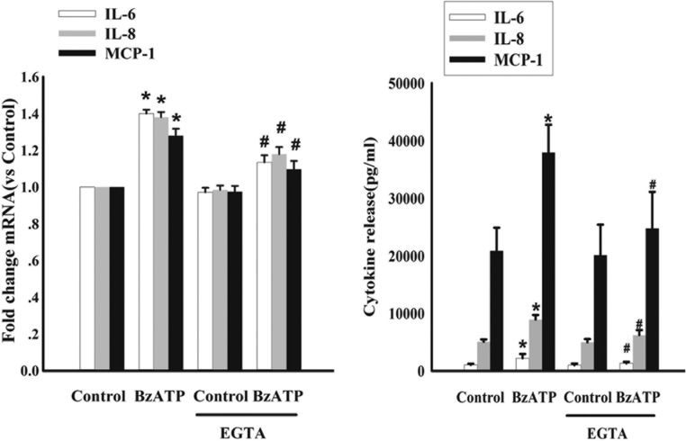
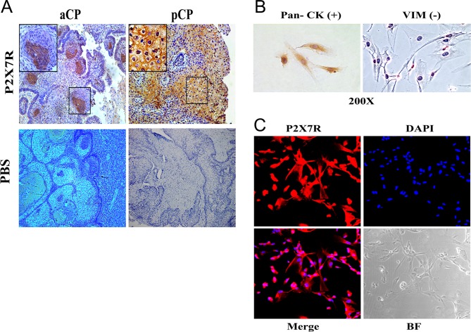
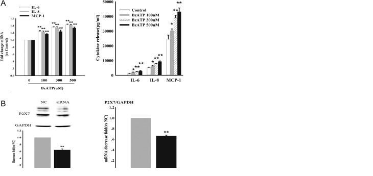
Craniopharyngiomas (CPs) are usually benign, non-metastasizing embryonic malformations originating from the sellar area. They are, however, locally invasive and generate adherent interfaces with the surrounding brain parenchyma. Previous studies have shown the tumor microenvironment is characterized by a local abundance of adenosine triphosphate (ATP), infiltration of leukocytes and elevated levels of pro-inflammatory cytokines that are thought to be responsible, at least in part, for the local invasion. Here, we examine whether ATP, via the P2X7R, participates in the regulation of cytokine expression in CPs. The expression of P2X7R and pro-inflammatory cytokines were measured at the RNA and protein levels both in tumor samples and in primary cultured tumor cells. Furthermore, cytokine modulation was measured after manipulating P2X7R in cultured tumor cells by siRNA-mediated knockdown, as well as pharmacologically by using selective agonists and antagonists. The following results were observed. A number of cytokines, in particular IL-6, IL-8 and MCP-1, were elevated in patient plasma, tumor tissue and cultured tumor cells. P2X7R was expressed in tumor tissue as well as in cultured tumor cells. RNA expression as measured in 48 resected tumors was positively correlated with the RNA levels of IL-6, IL-8 and MCP-1 in tumors. Furthermore, knockdown of P2X7R in primary tumor cultures reduced, and stimulation of P2XR7 by a specific agonist enhanced the expression of these cytokines. This latter stimulation involved a Ca-dependent mechanism and could be counteracted by the addition of an antagonist. In conclusion, the results suggest that P2X7R may promote IL-6, IL-8 and MCP-1 production and secretion and contribute to the invasion and adhesion of CPs to the surrounding tissue.
颅咽管瘤(CPs)通常是起源于鞍区的良性、非转移性胚胎畸形。然而,它们具有局部侵袭性,并与周围脑实质形成粘连界面。先前的研究表明,肿瘤微环境的特征是局部三磷酸腺苷(ATP)含量丰富、白细胞浸润以及促炎细胞因子水平升高,这些因素被认为至少部分地导致了局部侵袭。在此,我们研究ATP是否通过P2X7R参与颅咽管瘤中细胞因子表达的调控。我们在肿瘤样本和原代培养的肿瘤细胞中,从RNA和蛋白质水平检测了P2X7R和促炎细胞因子的表达。此外,我们通过小干扰RNA(siRNA)介导的敲低技术在培养的肿瘤细胞中操纵P2X7R,并使用选择性激动剂和拮抗剂进行药理学操作,然后检测细胞因子的调节情况。观察到以下结果。许多细胞因子,特别是白细胞介素-6(IL-6)、白细胞介素-8(IL-8)和单核细胞趋化蛋白-1(MCP-1),在患者血浆、肿瘤组织和培养的肿瘤细胞中均升高。P2X7R在肿瘤组织以及培养的肿瘤细胞中均有表达。在48个切除的肿瘤中检测到的RNA表达与肿瘤中IL-6、IL-8和MCP-1的RNA水平呈正相关。此外,原代肿瘤培养物中P2X7R的敲低降低了这些细胞因子的表达,而特异性激动剂对P2XR7的刺激则增强了这些细胞因子的表达。后者的刺激涉及一种钙依赖机制,并且可以通过添加拮抗剂来抵消。总之,结果表明P2X7R可能促进IL-6、IL-8和MCP-1的产生和分泌,并有助于颅咽管瘤向周围组织的侵袭和粘连。