• 文献检索
  • 文档翻译
  • 深度研究
  • 学术资讯
  • Suppr Zotero 插件Zotero 插件
  • 邀请有礼
  • 套餐&价格
  • 历史记录
应用&插件
Suppr Zotero 插件Zotero 插件浏览器插件Mac 客户端Windows 客户端微信小程序
定价
高级版会员购买积分包购买API积分包
服务
文献检索文档翻译深度研究API 文档MCP 服务
关于我们
关于 Suppr公司介绍联系我们用户协议隐私条款
关注我们

Suppr 超能文献

核心技术专利:CN118964589B侵权必究
粤ICP备2023148730 号-1Suppr @ 2026

文献检索

告别复杂PubMed语法,用中文像聊天一样搜索,搜遍4000万医学文献。AI智能推荐,让科研检索更轻松。

立即免费搜索

文件翻译

保留排版,准确专业,支持PDF/Word/PPT等文件格式,支持 12+语言互译。

免费翻译文档

深度研究

AI帮你快速写综述,25分钟生成高质量综述,智能提取关键信息,辅助科研写作。

立即免费体验

铜绿假单胞菌感染后角膜基质的二次谐波成像。

Second harmonic generation imaging of corneal stroma after infection by Pseudomonas aeruginosa.

机构信息

Department of Ophthalmology, The University of Texas Southwestern Medical Center, Dallas, TX, USA.

出版信息

Sci Rep. 2017 Apr 11;7:46116. doi: 10.1038/srep46116.

DOI:10.1038/srep46116
PMID:28397809
原文链接:https://pmc.ncbi.nlm.nih.gov/articles/PMC5387403/
Abstract

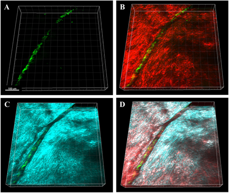
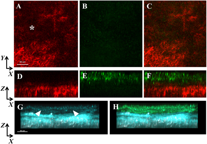
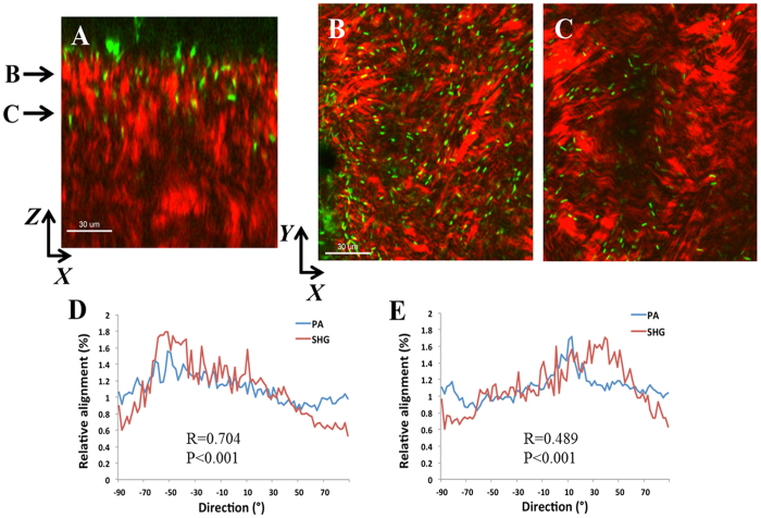
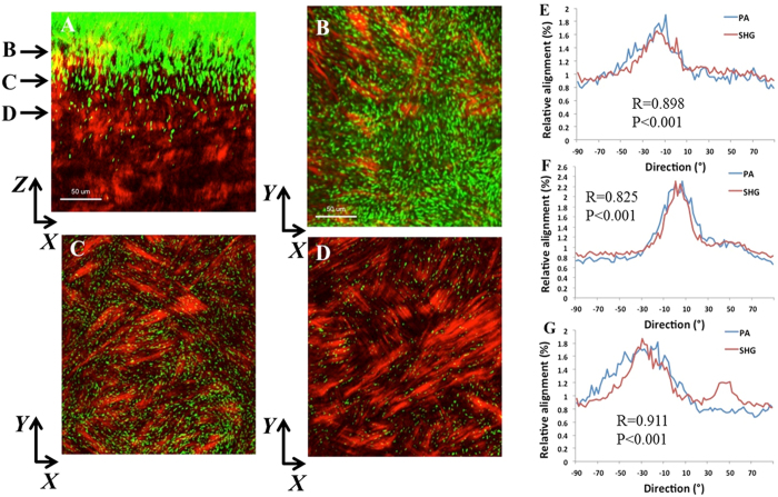
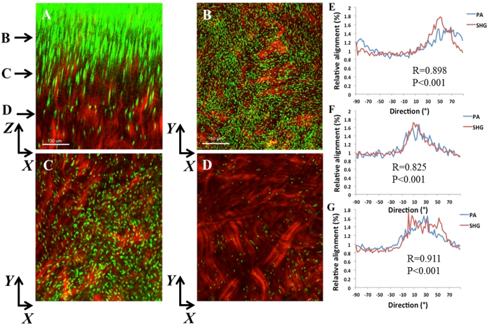
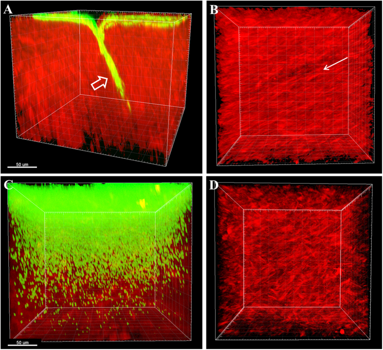
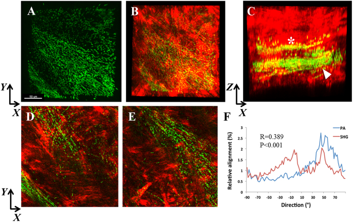
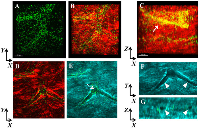
Pseudomonas aeruginosa is a pathogenic gram-negative organism that has the ability to cause blinding corneal infections following trauma and during contact lens wear. In this study, we investigated the directional movement and orientation of an invasive corneal isolate of P. aeruginosa in the corneal stroma during infection of ex vivo and in vivo rabbit corneas using multiphoton fluorescence and second harmonic generation (SHG) imaging. Ex vivo, rabbit corneas were subject to three partial thickness wounds prior to inoculation. In vivo, New Zealand white rabbits were fit with P. aeruginosa laden contact lenses in the absence of a penetrating wound. At all time points tested, infiltration of the corneal stroma by P. aeruginosa revealed a high degree of alignment between the bacteria and collagen lamellae ex vivo (p < 0.001). In vivo, P. aeruginosa traveled throughout the stroma in discrete regions or bands. Within each region, the bacteria showed good alignment with collagen lamellae (P = 0.002). Interestingly, in both the in vitro and in vivo models, P. aeruginosa did not appear to cross the corneal limbus. Taken together, our findings suggest that P. aeruginosa exploits the precise spacing of collagen lamellae in the central cornea to facilitate spread throughout the stroma.

摘要

铜绿假单胞菌是一种致病性革兰氏阴性菌,在创伤后和佩戴隐形眼镜时,具有引起致盲性角膜感染的能力。在这项研究中,我们使用多光子荧光和二次谐波产生(SHG)成像技术,研究了一种侵袭性角膜分离株铜绿假单胞菌在感染离体和体内兔角膜时在角膜基质中的定向运动和取向。离体时,在接种前,兔角膜经历了三个部分厚度的伤口。在体内,新西兰白兔在没有穿透性伤口的情况下佩戴载有铜绿假单胞菌的隐形眼镜。在所有测试的时间点,铜绿假单胞菌渗透到角膜基质中,显示出细菌与胶原板层之间高度的排列一致性(p < 0.001)。在体内,铜绿假单胞菌在离散区域或带中贯穿整个基质。在每个区域内,细菌与胶原板层排列良好(P = 0.002)。有趣的是,在体外和体内模型中,铜绿假单胞菌似乎都没有穿过角膜缘。总之,我们的发现表明,铜绿假单胞菌利用中央角膜中胶原板层的精确间距来促进其在基质中的传播。

https://cdn.ncbi.nlm.nih.gov/pmc/blobs/6daf/5387403/0ffcc481c581/srep46116-f8.jpg
https://cdn.ncbi.nlm.nih.gov/pmc/blobs/6daf/5387403/29673433ced4/srep46116-f1.jpg
https://cdn.ncbi.nlm.nih.gov/pmc/blobs/6daf/5387403/9f1956df4eba/srep46116-f2.jpg
https://cdn.ncbi.nlm.nih.gov/pmc/blobs/6daf/5387403/a8cc77f18843/srep46116-f3.jpg
https://cdn.ncbi.nlm.nih.gov/pmc/blobs/6daf/5387403/1d3cee574976/srep46116-f4.jpg
https://cdn.ncbi.nlm.nih.gov/pmc/blobs/6daf/5387403/f2d93fb6d164/srep46116-f5.jpg
https://cdn.ncbi.nlm.nih.gov/pmc/blobs/6daf/5387403/079a1481e090/srep46116-f6.jpg
https://cdn.ncbi.nlm.nih.gov/pmc/blobs/6daf/5387403/9c7996b0eb8b/srep46116-f7.jpg
https://cdn.ncbi.nlm.nih.gov/pmc/blobs/6daf/5387403/0ffcc481c581/srep46116-f8.jpg

相似文献

1
Second harmonic generation imaging of corneal stroma after infection by Pseudomonas aeruginosa.铜绿假单胞菌感染后角膜基质的二次谐波成像。
Sci Rep. 2017 Apr 11;7:46116. doi: 10.1038/srep46116.
2
A partial-thickness epithelial defect increases the adherence of Pseudomonas aeruginosa to the cornea.部分厚度上皮缺损会增加铜绿假单胞菌对角膜的黏附。
Invest Ophthalmol Vis Sci. 1989 Jun;30(6):1069-74.
3
Prolonged hypoxia induces lipid raft formation and increases Pseudomonas internalization in vivo after contact lens wear and lid closure.长时间缺氧会诱导脂筏形成,并增加佩戴隐形眼镜和闭眼后体内铜绿假单胞菌的内化。
Eye Contact Lens. 2006 May;32(3):114-20. doi: 10.1097/01.icl.0000177384.27778.4c.
4
Multiphoton microscopy of ex vivo corneas after collagen cross-linking.胶原交联术后离体角膜的多光子显微镜检查。
Invest Ophthalmol Vis Sci. 2011 Jul 18;52(8):5325-31. doi: 10.1167/iovs.11-7184.
5
Different strains of Pseudomonas aeruginosa isolated from ocular infections or inflammation display distinct corneal pathologies in an animal model.从眼部感染或炎症中分离出的不同铜绿假单胞菌菌株在动物模型中表现出不同的角膜病变。
Curr Eye Res. 1998 Jul;17(7):730-5.
6
[Adherence of Pseudomonas aeruginosa to the rabbit corneal epithelium].[铜绿假单胞菌对兔角膜上皮的黏附]
Nippon Ganka Gakkai Zasshi. 1990 Mar;94(3):269-76.
7
Internalization of Pseudomonas aeruginosa is mediated by lipid rafts in contact lens-wearing rabbit and cultured human corneal epithelial cells.铜绿假单胞菌的内化作用是由佩戴隐形眼镜的兔和培养的人角膜上皮细胞中的脂筏介导的。
Invest Ophthalmol Vis Sci. 2005 Apr;46(4):1348-55. doi: 10.1167/iovs.04-0542.
8
Microbiologic, Pharmacokinetic, and Clinical Effects of Corneal Collagen Cross-Linking on Experimentally Induced Pseudomonas Keratitis in Rabbits.角膜胶原交联对兔实验性铜绿假单胞菌角膜炎的微生物学、药代动力学及临床效应
Cornea. 2015 Oct;34(10):1276-80. doi: 10.1097/ICO.0000000000000516.
9
Adherence of Pseudomonas aeruginosa to shed rabbit corneal epithelial cells after overnight wear of contact lenses.过夜佩戴隐形眼镜后铜绿假单胞菌对脱落兔角膜上皮细胞的黏附
CLAO J. 1997 Jan;23(1):63-8.
10
Corneal Fibroblast Migration Patterns During Intrastromal Wound Healing Correlate With ECM Structure and Alignment.基质内伤口愈合过程中角膜成纤维细胞迁移模式与细胞外基质结构及排列相关。
Invest Ophthalmol Vis Sci. 2015 Nov;56(12):7352-61. doi: 10.1167/iovs.15-17978.

引用本文的文献

1
Two-Photon Imaging for Non-Invasive Corneal Examination.双光子成像技术在非侵入性角膜检查中的应用。
Sensors (Basel). 2022 Dec 11;22(24):9699. doi: 10.3390/s22249699.
2
Genipin in an Ex Vivo Corneal Model of Bacterial and Fungal Keratitis.金皮啶在细菌性和真菌性角膜炎的离体角膜模型中的作用。
Transl Vis Sci Technol. 2021 Aug 2;10(9):31. doi: 10.1167/tvst.10.9.31.
3
Cationic Polyelectrolyte Nanocapsules of Moxifloxacin for Microbial Keratitis Therapy: Development, Characterization, and Pharmacodynamic Study.莫西沙星阳离子聚电解质纳米胶囊用于微生物角膜炎治疗:开发、表征和药效学研究。

本文引用的文献

1
Comparison of reflectance confocal microscopy and two-photon second harmonic generation microscopy in fungal keratitis rabbit model ex vivo.反射共聚焦显微镜与双光子二次谐波产生显微镜在真菌性角膜炎兔离体模型中的比较
Biomed Opt Express. 2016 Jan 26;7(2):677-87. doi: 10.1364/BOE.7.000677. eCollection 2016 Feb 1.
2
Corneal Fibroblast Migration Patterns During Intrastromal Wound Healing Correlate With ECM Structure and Alignment.基质内伤口愈合过程中角膜成纤维细胞迁移模式与细胞外基质结构及排列相关。
Invest Ophthalmol Vis Sci. 2015 Nov;56(12):7352-61. doi: 10.1167/iovs.15-17978.
3
Corneal structure and transparency.
AAPS PharmSciTech. 2021 Jun 28;22(5):195. doi: 10.1208/s12249-021-02039-1.
4
An Organ System-Based Synopsis of Virulence.基于器官系统的毒力概要
Virulence. 2021 Dec;12(1):1469-1507. doi: 10.1080/21505594.2021.1926408.
5
Corneal Infection Models: Tools to Investigate the Role of Biofilms in Bacterial Keratitis.角膜感染模型:研究生物膜在细菌性角膜炎中作用的工具。
Cells. 2020 Nov 10;9(11):2450. doi: 10.3390/cells9112450.
6
An cornea infection model.一种角膜感染模型。 (注:原英文有误,正确应为“A corneal infection model” )
MethodsX. 2020 Apr 7;7:100876. doi: 10.1016/j.mex.2020.100876. eCollection 2020.
7
Contact lens-related corneal infection: Intrinsic resistance and its compromise.接触镜相关的角膜感染:固有抵抗力及其受损。
Prog Retin Eye Res. 2020 May;76:100804. doi: 10.1016/j.preteyeres.2019.100804. Epub 2019 Nov 20.
8
Quantitative Discrimination of Healthy and Diseased Corneas With Second Harmonic Generation Microscopy.利用二次谐波产生显微镜对健康角膜和患病角膜进行定量鉴别。
Transl Vis Sci Technol. 2019 Jun 27;8(3):51. doi: 10.1167/tvst.8.3.51. eCollection 2019 May.
角膜结构与透明度。
Prog Retin Eye Res. 2015 Nov;49:1-16. doi: 10.1016/j.preteyeres.2015.07.001. Epub 2015 Jul 2.
4
From nano to macro: studying the hierarchical structure of the corneal extracellular matrix.从纳米到宏观:研究角膜细胞外基质的层级结构
Exp Eye Res. 2015 Apr;133:81-99. doi: 10.1016/j.exer.2014.07.018.
5
Quantitative analysis of collagen lamellae in the normal and keratoconic human cornea by second harmonic generation imaging microscopy.通过二次谐波产生成像显微镜对正常和圆锥角膜人眼角膜中的胶原板层进行定量分析。
Invest Ophthalmol Vis Sci. 2014 Nov 25;55(12):8377-85. doi: 10.1167/iovs.14-15348.
6
Pseudomonas aeruginosa infectious keratitis in a high oxygen transmissible rigid contact lens rabbit model.高氧通透性硬性角膜接触镜兔模型中的铜绿假单胞菌感染性角膜炎。
Invest Ophthalmol Vis Sci. 2014 Aug 14;55(9):5890-9. doi: 10.1167/iovs.14-14235.
7
Prevailing role of contact guidance in intrastromal T-cell trapping in human pancreatic cancer.接触引导在人胰腺癌细胞间质内 T 细胞捕获中的普遍作用。
Clin Cancer Res. 2014 Jul 1;20(13):3422-33. doi: 10.1158/1078-0432.CCR-13-2972. Epub 2014 Apr 24.
8
Three-dimensional distribution of transverse collagen fibers in the anterior human corneal stroma.人眼角膜前部基质中横向胶原纤维的三维分布。
Invest Ophthalmol Vis Sci. 2013 Nov 5;54(12):7293-301. doi: 10.1167/iovs.13-13150.
9
The ocular surface epithelial barrier and other mechanisms of mucosal protection: from allergy to infectious diseases.眼表面上皮屏障和其他黏膜保护机制:从过敏到传染病。
Curr Opin Allergy Clin Immunol. 2013 Oct;13(5):563-8. doi: 10.1097/ACI.0b013e3283645899.
10
Cytokeratins mediate epithelial innate defense through their antimicrobial properties.细胞角蛋白通过其抗菌特性介导上皮固有防御。
J Clin Invest. 2012 Oct;122(10):3665-77. doi: 10.1172/JCI64416. Epub 2012 Sep 24.