• 文献检索
  • 文档翻译
  • 深度研究
  • 学术资讯
  • Suppr Zotero 插件Zotero 插件
  • 邀请有礼
  • 套餐&价格
  • 历史记录
应用&插件
Suppr Zotero 插件Zotero 插件浏览器插件Mac 客户端Windows 客户端微信小程序
定价
高级版会员购买积分包购买API积分包
服务
文献检索文档翻译深度研究API 文档MCP 服务
关于我们
关于 Suppr公司介绍联系我们用户协议隐私条款
关注我们

Suppr 超能文献

核心技术专利:CN118964589B侵权必究
粤ICP备2023148730 号-1Suppr @ 2026

文献检索

告别复杂PubMed语法,用中文像聊天一样搜索,搜遍4000万医学文献。AI智能推荐,让科研检索更轻松。

立即免费搜索

文件翻译

保留排版,准确专业,支持PDF/Word/PPT等文件格式,支持 12+语言互译。

免费翻译文档

深度研究

AI帮你快速写综述,25分钟生成高质量综述,智能提取关键信息,辅助科研写作。

立即免费体验

脊髓小脑共济失调3型和10型的光学相干断层扫描对比研究

A Comparative Optical Coherence Tomography Study of Spinocerebellar Ataxia Types 3 and 10.

作者信息

Spina Tensini Fernando, Sato Mario T, Shiokawa Naoye, Ashizawa Tetsuo, Teive Hélio A G

机构信息

Movement Disorders Unit, Neurology Service, Internal Medicine Department, Hospital de Clínicas, Federal University of Paraná, Rua Treze de Maio, 200, apto 103, Centro, Curitiba, PR, CEP 80020-270, Brazil.

Neuro-Ophthalmology and Ocular Electrophysiology Sector, Vision Center, Hospital de Clínicas, Federal University of Paraná, Curitiba, PR, Brazil.

出版信息

Cerebellum. 2017 Aug;16(4):797-801. doi: 10.1007/s12311-017-0856-7.

DOI:10.1007/s12311-017-0856-7
PMID:28401494
原文链接:https://pmc.ncbi.nlm.nih.gov/articles/PMC6996148/
Abstract

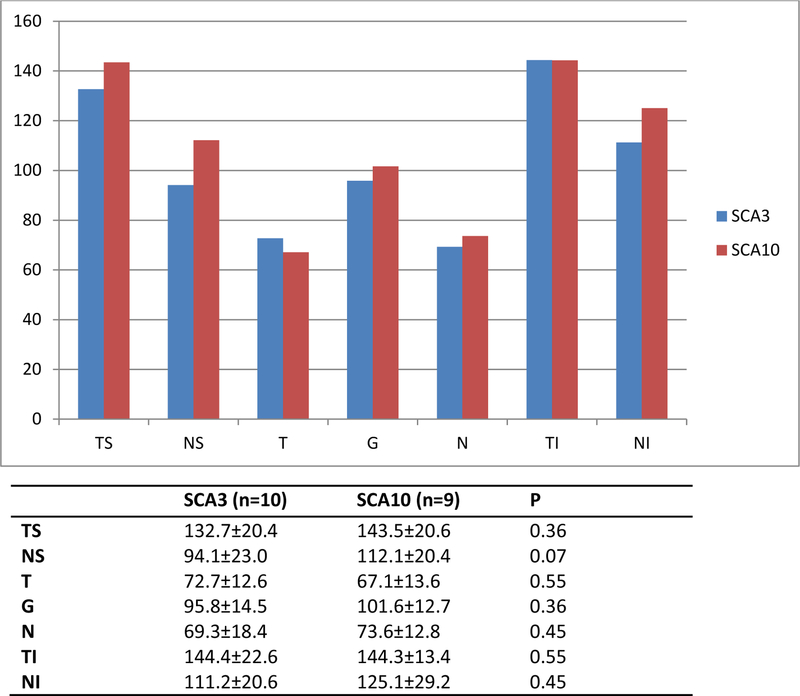
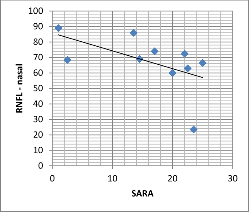
SCA3 presents with a CAG expansion at 14q24.3-q32 while SCA10 shows an ATTCT expansion at 22q13-qter. SCA10 seems to be less aggressive than SCA3. For an in vivo, noninvasive approach of the correlation between central nervous system and clinical evolution, we can use optic coherence tomography (OCT) to measure retinal nerve fiber (RNFL) and ganglion cell layer (GCL) thickness. To describe OCT findings in SCA10, correlate it with expansion size and disease severity and compare with those of SCA3. We analyzed ten individuals with SCA3 and nine with SCA10 recruited from the neurology service of Hospital de Clínicas of Paraná-Brazil. They were submitted to OCT and clinical evaluation using SARA score. Expansion size, demographic data, time from disease onset, and age of onset were collected. We found no correlation between size of expansion, SARA, and RNFL or GCL thickness in SCA10. RNFL seemed to be thicker in SCA10 (p > 0.05). GCL thickness, SARA, median age, and time from disease onset did not differ between groups. SCA10 individuals had an earlier disease onset. In SCA3, there was a negative correlation between SARA and RNFL thickness in nasal area. To the best of our knowledge, this is the first paper assessing retinal changes by OCT in individuals with SCA10. The lack of correlation between disease progression, age, and time since onset supports the anatomopathological findings which suggest SCA10 is less aggressive than other SCAs. The findings in SCA3 are in accordance with the literature.

摘要

脊髓小脑共济失调3型(SCA3)表现为14q24.3 - q32处的CAG重复序列扩增,而脊髓小脑共济失调10型(SCA10)则显示22q13 - qter处的ATTCT重复序列扩增。SCA10似乎比SCA3的侵袭性小。为了采用一种体内非侵入性方法研究中枢神经系统与临床进展之间的相关性,我们可以使用光学相干断层扫描(OCT)来测量视网膜神经纤维层(RNFL)和神经节细胞层(GCL)的厚度。为了描述SCA10患者的OCT检查结果,将其与重复序列扩增大小和疾病严重程度相关联,并与SCA3患者的结果进行比较。我们分析了从巴西巴拉那州临床医院神经科招募的10例SCA3患者和9例SCA10患者。他们接受了OCT检查,并使用共济失调评定量表(SARA)进行临床评估。收集了重复序列扩增大小、人口统计学数据、疾病发病时间和发病年龄。我们发现SCA10患者的重复序列扩增大小、SARA评分与RNFL或GCL厚度之间无相关性。SCA10患者的RNFL似乎更厚(p > 0.05)。两组之间的GCL厚度、SARA评分、中位年龄和疾病发病时间无差异。SCA10患者的发病年龄较早。在SCA3患者中,SARA评分与鼻侧RNFL厚度呈负相关。据我们所知,这是第一篇评估SCA10患者视网膜变化的OCT研究论文。疾病进展、年龄和发病时间之间缺乏相关性,支持了解剖病理学研究结果,即SCA10比其他脊髓小脑共济失调的侵袭性小。SCA3的研究结果与文献一致。

https://cdn.ncbi.nlm.nih.gov/pmc/blobs/b01c/6996148/5c2c7de05d91/nihms-1066357-f0002.jpg
https://cdn.ncbi.nlm.nih.gov/pmc/blobs/b01c/6996148/c07f950670e9/nihms-1066357-f0001.jpg
https://cdn.ncbi.nlm.nih.gov/pmc/blobs/b01c/6996148/5c2c7de05d91/nihms-1066357-f0002.jpg
https://cdn.ncbi.nlm.nih.gov/pmc/blobs/b01c/6996148/c07f950670e9/nihms-1066357-f0001.jpg
https://cdn.ncbi.nlm.nih.gov/pmc/blobs/b01c/6996148/5c2c7de05d91/nihms-1066357-f0002.jpg

相似文献

1
A Comparative Optical Coherence Tomography Study of Spinocerebellar Ataxia Types 3 and 10.脊髓小脑共济失调3型和10型的光学相干断层扫描对比研究
Cerebellum. 2017 Aug;16(4):797-801. doi: 10.1007/s12311-017-0856-7.
2
Volumetric MRI Changes in Spinocerebellar Ataxia (SCA3 and SCA10) Patients.小脑脊髓性共济失调(SCA3 和 SCA10)患者的容积 MRI 变化。
Cerebellum. 2020 Aug;19(4):536-543. doi: 10.1007/s12311-020-01137-3.
3
Analysis of diffusion tensor parameters in spinocerebellar ataxia type 3 and type 10 patients.脊髓小脑性共济失调 3 型和 10 型患者的弥散张量参数分析。
Parkinsonism Relat Disord. 2020 Sep;78:73-78. doi: 10.1016/j.parkreldis.2020.06.460. Epub 2020 Jul 27.
4
Characterization of Retinal Architecture in Spinocerebellar Ataxia Type 3 and Correlation with Disease Severity.脊髓小脑共济失调 3 型的视网膜结构特征及其与疾病严重程度的相关性。
Mov Disord. 2022 Apr;37(4):758-766. doi: 10.1002/mds.28893. Epub 2021 Dec 22.
5
Progression of Retinal Ganglion Cell and Nerve Fiber Layer Loss in Spinocerebellar Ataxia 3 Patients.脊髓小脑性共济失调 3 型患者的视网膜神经节细胞和神经纤维层丢失进展。
Cerebellum. 2024 Aug;23(4):1348-1354. doi: 10.1007/s12311-023-01634-1. Epub 2023 Nov 30.
6
Body composition in Spinocerebellar ataxia type 3 and 10 patients: Comparative study with control group.3 型和 10 型脊髓小脑共济失调患者的身体成分:与对照组的对比研究。
Nutr Neurosci. 2020 Jan;23(1):49-54. doi: 10.1080/1028415X.2018.1469282. Epub 2018 May 7.
7
Olfactory Function in SCA10.SCA10 中的嗅觉功能。
Cerebellum. 2019 Feb;18(1):85-90. doi: 10.1007/s12311-018-0954-1.
8
Comparing loss of balance and functional capacity among patients with SCA2, SCA3 and SCA10.比较SCA2、SCA3和SCA10患者的平衡能力丧失和功能能力。
Clin Neurol Neurosurg. 2022 Mar;214:107150. doi: 10.1016/j.clineuro.2022.107150. Epub 2022 Feb 1.
9
Spinocerebellar ataxia in a cohort of patients from Rio de Janeiro.来自里约热内卢的一组患者的脊髓小脑共济失调。
Neurol Sci. 2022 Aug;43(8):4997-5005. doi: 10.1007/s10072-022-06084-x. Epub 2022 Apr 25.
10
Conversion of individuals at risk for spinocerebellar ataxia types 1, 2, 3, and 6 to manifest ataxia (RISCA): a longitudinal cohort study.将脊髓小脑共济失调 1、2、3、6 型的风险个体转化为显性共济失调(RISCA):一项纵向队列研究。
Lancet Neurol. 2020 Sep;19(9):738-747. doi: 10.1016/S1474-4422(20)30235-0.

引用本文的文献

1
Progression of Retinal Ganglion Cell and Nerve Fiber Layer Loss in Spinocerebellar Ataxia 3 Patients.脊髓小脑性共济失调 3 型患者的视网膜神经节细胞和神经纤维层丢失进展。
Cerebellum. 2024 Aug;23(4):1348-1354. doi: 10.1007/s12311-023-01634-1. Epub 2023 Nov 30.
2
The genetic and molecular features of the intronic pentanucleotide repeat expansion in spinocerebellar ataxia type 10.10型脊髓小脑共济失调中内含子五核苷酸重复扩增的遗传和分子特征
Front Genet. 2022 Sep 15;13:936869. doi: 10.3389/fgene.2022.936869. eCollection 2022.
3
Reply to: "Microvascular Breakdown Due to Retinal Neurodegeneration in Ataxias".

本文引用的文献

1
Ganglion cell layer measurements correlate with disease severity in patients with Alzheimer's disease.在阿尔茨海默病患者中,神经节细胞层测量结果与疾病严重程度相关。
Acta Ophthalmol. 2016 Sep;94(6):e454-9. doi: 10.1111/aos.12977. Epub 2016 Feb 19.
2
Optical Coherence Tomography for the Detection of Remote Optic Neuritis in Multiple Sclerosis.光学相干断层扫描用于检测多发性硬化症中的远隔性视神经炎
J Neuroimaging. 2016 May;26(3):283-8. doi: 10.1111/jon.12326. Epub 2016 Jan 15.
3
Optical coherence tomography as a tool to evaluate retinal changes in Parkinson's disease.
回复:“共济失调中视网膜神经变性导致的微血管破裂”
Mov Disord. 2022 Feb;37(2):438. doi: 10.1002/mds.28916. Epub 2022 Jan 10.
4
Zebrafish Models of Autosomal Dominant Ataxias.常染色体显性遗传性共济失调症的斑马鱼模型。
Cells. 2021 Feb 17;10(2):421. doi: 10.3390/cells10020421.
5
The Deubiquitinating Enzyme Ataxin-3 Regulates Ciliogenesis and Phagocytosis in the Retina.去泛素化酶 Ataxin-3 调控视网膜中的纤毛发生和吞噬作用。
Cell Rep. 2020 Nov 10;33(6):108360. doi: 10.1016/j.celrep.2020.108360.
6
Ophthalmic Manifestations and Genetics of the Polyglutamine Autosomal Dominant Spinocerebellar Ataxias: A Review.多聚谷氨酰胺常染色体显性遗传性脊髓小脑共济失调的眼科表现与遗传学:综述
Front Neurosci. 2020 Aug 21;14:892. doi: 10.3389/fnins.2020.00892. eCollection 2020.
7
Dystonia in Patients with Spinocerebellar Ataxia 3 - Machado-Joseph disease: An Underestimated Diagnosis?脊髓小脑共济失调3型-马查多-约瑟夫病患者的肌张力障碍:一个被低估的诊断?
Open Neurol J. 2018 May 31;12:41-49. doi: 10.2174/1874205X01812010041. eCollection 2018.
光学相干断层扫描作为评估帕金森病视网膜变化的工具。
Parkinsonism Relat Disord. 2015 Oct;21(10):1164-9. doi: 10.1016/j.parkreldis.2015.08.002. Epub 2015 Aug 10.
4
Primary and secondary ataxias.原发性和继发性共济失调。
Curr Opin Neurol. 2015 Aug;28(4):413-22. doi: 10.1097/WCO.0000000000000227.
5
Mutational screening of 320 Brazilian patients with autosomal dominant spinocerebellar ataxia.对320名患有常染色体显性遗传性脊髓小脑共济失调的巴西患者进行突变筛查。
J Neurol Sci. 2014 Dec 15;347(1-2):375-9. doi: 10.1016/j.jns.2014.10.036. Epub 2014 Oct 31.
6
Retinal neurodegeneration on optical coherence tomography and cerebral atrophy.光学相干断层扫描显示的视网膜神经变性和脑萎缩。
Neurosci Lett. 2015 Jan 1;584:12-6. doi: 10.1016/j.neulet.2014.10.010. Epub 2014 Oct 19.
7
Relationship Between Optic Nerve Appearance and Retinal Nerve Fiber Layer Thickness as Explored with Spectral Domain Optical Coherence Tomography.利用光谱域光学相干断层扫描技术探究视神经外观与视网膜神经纤维层厚度之间的关系
Transl Vis Sci Technol. 2014 Nov 3;3(6):4. doi: 10.1167/tvst.3.6.4. eCollection 2014 Oct.
8
Genetic regulation of vertebrate eye development.脊椎动物眼睛发育的遗传调控。
Clin Genet. 2014 Nov;86(5):453-60. doi: 10.1111/cge.12493. Epub 2014 Sep 25.
9
Spinocerebellar ataxia type 10 in Peru: the missing link in the Amerindian origin of the disease.秘鲁的10型脊髓小脑共济失调:该疾病美洲印第安人起源中缺失的环节。
J Neurol. 2014 Sep;261(9):1691-4. doi: 10.1007/s00415-014-7394-8. Epub 2014 Jun 17.
10
The association of spectral-domain optical coherence tomography determined ganglion cell complex parameters and disease severity in Parkinson's disease.频域光学相干断层扫描测定的帕金森病神经节细胞复合体参数与疾病严重程度的关联。
Curr Eye Res. 2014 Nov;39(11):1117-22. doi: 10.3109/02713683.2014.894080. Epub 2014 Mar 21.