Taylor Erik N, Ding Yao, Zhu Shan, Cheah Eric, Alexander Phillip, Lin Leon, Aninwene George E, Hoffman Matthew P, Mahajan Anita, Mohamed Abdallah S R, McDannold Nathan, Fuller Clifton D, Chen Clark C, Gilbert Richard J
Chemistry and Chemical Biology, Northeastern University, Boston, MA, USA.
Radiation Oncology, University of Texas, MD Anderson Cancer Center, Houston, TX, USA.
Oncotarget. 2017 Jun 27;8(26):41815-41826. doi: 10.18632/oncotarget.16296.
While it is recognized that the overall resistance of glioblastoma to treatment may be related to intra-tumor patterns of structural heterogeneity, imaging methods to assess such patterns remain rudimentary.
We utilized a generalized Q-space imaging (GQI) algorithm to analyze magnetic resonance imaging (MRI) derived from a rodent model of glioblastoma and 2 clinical datasets to correlate GQI, histology, and survival.
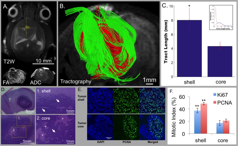
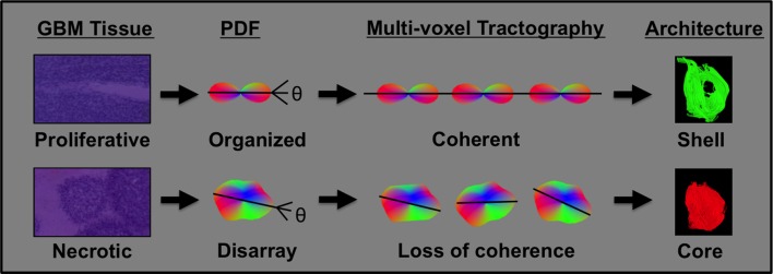
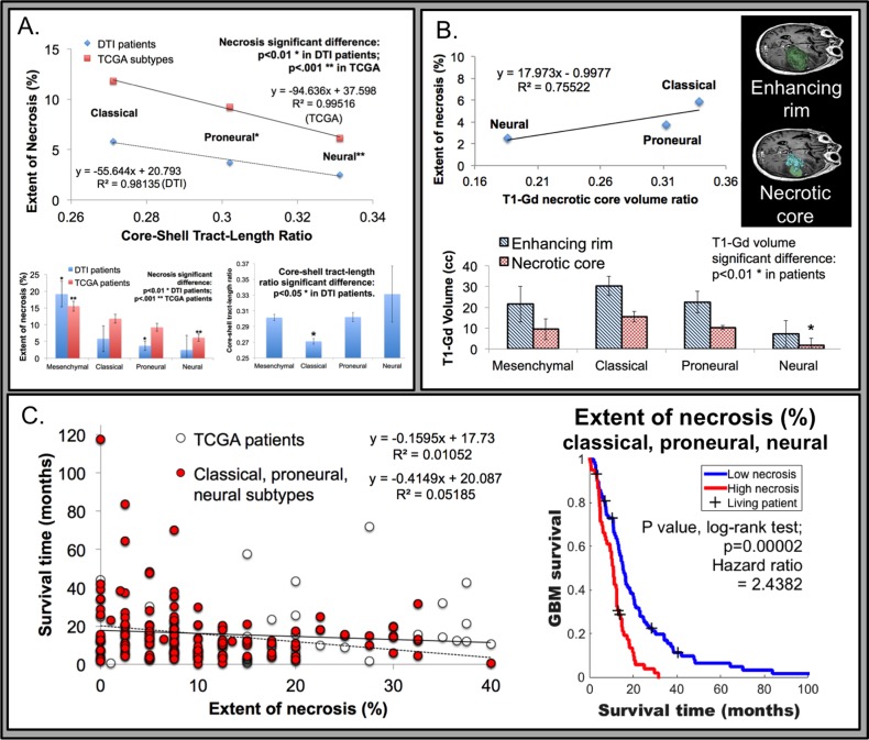
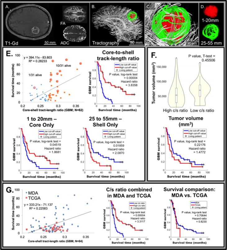
In a rodent glioblastoma model, GQI demonstrated a poorly coherent core region, consisting of diffusion tracts <5 mm, surrounded by a shell of highly coherent diffusion tracts, 6-25 mm. Histologically, the core region possessed a high degree of necrosis, whereas the shell consisted of organized sheets of anaplastic cells with elevated mitotic index. These attributes define tumor architecture as the macroscopic organization of variably aligned tumor cells. Applied to MRI data from The Cancer Imaging Atlas (TCGA), the core-shell diffusion tract-length ratio (c/s ratio) correlated linearly with necrosis, which, in turn, was inversely associated with survival (p = 0.00002). We confirmed in an independent cohort of patients (n = 62) that the c/s ratio correlated inversely with survival (p = 0.0004).
The analysis of MR images by GQI affords insight into tumor architectural patterns in glioblastoma that correlate with biological heterogeneity and clinical outcome.
虽然人们认识到胶质母细胞瘤对治疗的总体抗性可能与肿瘤内结构异质性模式有关,但评估此类模式的成像方法仍很初级。
我们利用广义Q空间成像(GQI)算法分析源自胶质母细胞瘤啮齿动物模型的磁共振成像(MRI)以及两个临床数据集,以关联GQI、组织学和生存率。
在一个啮齿动物胶质母细胞瘤模型中,GQI显示出一个连贯性较差的核心区域,由长度小于5毫米的扩散束组成,周围是一个由长度为6 - 25毫米的高度连贯扩散束构成的壳层。组织学上,核心区域具有高度坏死,而壳层由有丝分裂指数升高的间变性细胞有序片层组成。这些特征将肿瘤结构定义为不同排列的肿瘤细胞的宏观组织。应用于来自癌症成像图谱(TCGA)的MRI数据时,核心 - 壳层扩散束长度比(c/s比)与坏死呈线性相关,而坏死又与生存率呈负相关(p = 0.00002)。我们在一个独立的患者队列(n = 62)中证实,c/s比与生存率呈负相关(p = 0.0004)。
通过GQI分析MR图像能够深入了解胶质母细胞瘤的肿瘤结构模式,这些模式与生物学异质性和临床结果相关。