Youssofzadeh Vahab, Williamson Brady J, Kadis Darren S
Pediatric Neuroimaging Research Consortium (PNRC), Cincinnati Children's Hospital Medical CenterCincinnati, OH, USA.
Division of Neurology, Cincinnati Children's Hospital Medical CenterCincinnati, OH, USA.
Front Hum Neurosci. 2017 Apr 5;11:173. doi: 10.3389/fnhum.2017.00173. eCollection 2017.
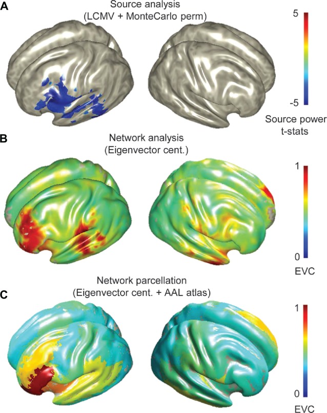
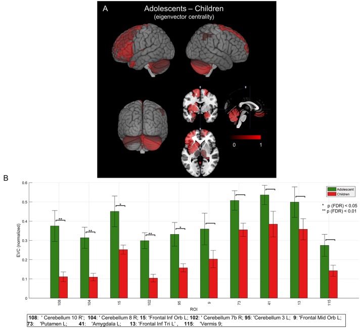
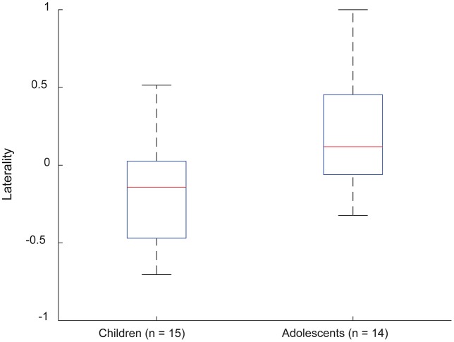
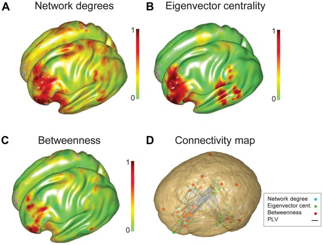
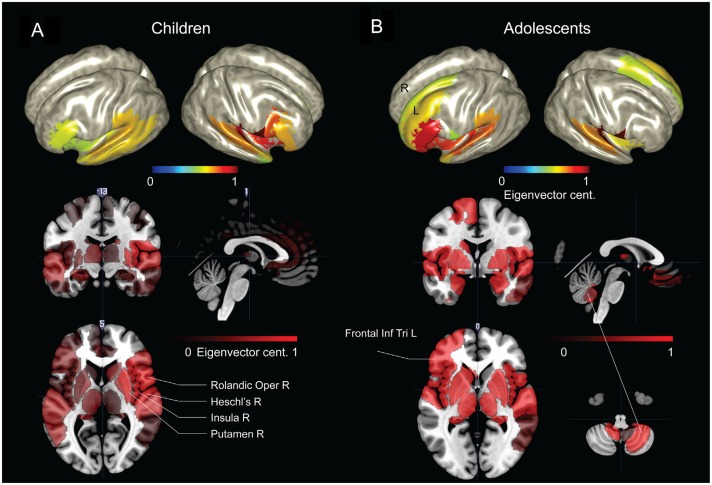
A classic left frontal-temporal brain network is known to support language processes. However, the level of participation of constituent regions, and the contribution of extra-canonical areas, is not fully understood; this is particularly true in children, and in individuals who have experienced early neurological insult. In the present work, we propose whole-brain connectivity and graph-theoretical analysis of magnetoencephalography (MEG) source estimates to provide robust maps of the pediatric expressive language network. We examined neuromagnetic data from a group of typically-developing young children ( = 15, ages 4-6 years) and adolescents ( = 14, 16-18 years) completing an auditory verb generation task in MEG. All source analyses were carried out using a linearly-constrained minimum-variance (LCMV) beamformer. Conventional differential analyses revealed significant ( < 0.05, corrected) low-beta (13-23 Hz) event related desynchrony (ERD) focused in the left inferior frontal region (Broca's area) in both groups, consistent with previous studies. Connectivity analyses were carried out in broadband (3-30 Hz) on time-course estimates obtained at the voxel level. Patterns of connectivity were characterized by (PLV), and network hubs identified through . Hub analysis revealed the importance of left perisylvian sites, i.e., Broca's and Wernicke's areas, across groups. The hemispheric distribution of frontal and temporal lobe EVC values was asymmetrical in most subjects; left dominant EVC was observed in 20% of young children, and 71% of adolescents. Interestingly, the adolescent group demonstrated increased critical sites in the right cerebellum, left inferior frontal gyrus (IFG) and left putamen. Here, we show that whole brain connectivity and network analysis can be used to map critical language sites in typical development; these methods may be useful for defining the margins of eloquent tissue in neurosurgical candidates.
已知一个经典的左额颞脑网络支持语言处理过程。然而,组成区域的参与程度以及非典型区域的贡献尚未完全明确;在儿童以及经历过早发性神经损伤的个体中尤其如此。在本研究中,我们提出对脑磁图(MEG)源估计进行全脑连通性和图论分析,以提供小儿表达性语言网络的可靠图谱。我们检查了一组在MEG中完成听觉动词生成任务的发育正常的幼儿(n = 15,4 - 6岁)和青少年(n = 14,16 - 18岁)的神经磁数据。所有源分析均使用线性约束最小方差(LCMV)波束形成器进行。传统的差异分析显示,两组在左额下回区域(布洛卡区)均出现显著(p < 0.05,校正)的低β频段(13 - 23 Hz)事件相关去同步化(ERD),这与先前的研究一致。连通性分析在宽带(3 - 30 Hz)上对体素水平获得的时间进程估计值进行。连通性模式通过相位锁定值(PLV)进行表征,并通过聚类系数识别网络枢纽。枢纽分析揭示了跨组的左外侧裂周围区域(即布洛卡区和韦尼克区)的重要性。大多数受试者额叶和颞叶等效视野(EVC)值的半球分布不对称;20%的幼儿和71%的青少年观察到左侧优势EVC。有趣的是,青少年组在右侧小脑、左侧额下回(IFG)和左侧壳核中显示出关键位点增加。在这里,我们表明全脑连通性和网络分析可用于绘制典型发育中的关键语言位点;这些方法可能有助于确定神经外科手术候选者中明确组织的边界。