Lee Yubu, Park Bo-Yong, James Oliver, Kim Seong-Gi, Park Hyunjin
Center for Neuroscience Imaging Research, Institute for Basic Science (IBS)Suwon, South Korea.
Department of Electronic, Electrical and Computer Engineering, Sungkyunkwan UniversitySuwon, South Korea.
Front Hum Neurosci. 2017 Aug 18;11:418. doi: 10.3389/fnhum.2017.00418. eCollection 2017.
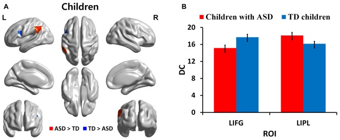
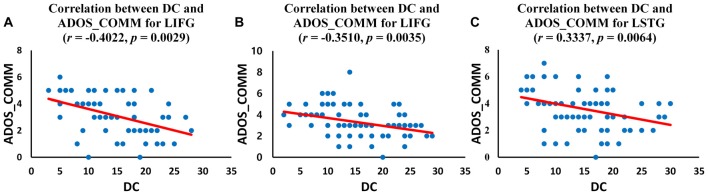
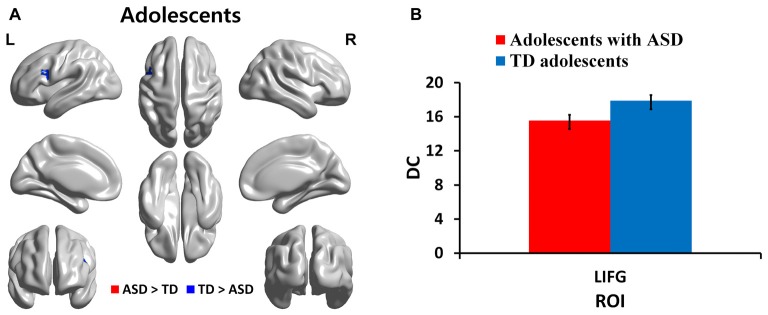
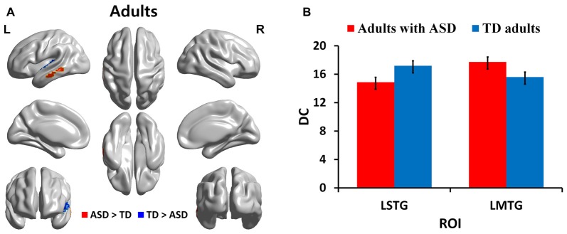
Autism spectrum disorder (ASD) is a neurodevelopmental disability with global implication. Altered brain connectivity in the language network has frequently been reported in ASD patients using task-based functional magnetic resonance imaging (fMRI) compared to typically developing (TD) participants. Most of these studies have focused on a specific age group or mixed age groups with ASD. In the current study, we investigated age-related changes in functional connectivity related measure, degree centrality (DC), in the language network across three age groups with ASD (113 children, 113 adolescents and 103 adults) using resting-state fMRI data collected from the autism brain imaging data exchange repository. We identified regions with significant group-wise differences between ASD and TD groups for three age cohorts using DC based on graph theory. We found that both children and adolescents with ASD showed decreased DC in Broca's area compared to age-matched TD groups. Adults with ASD showed decreased DC in Wernicke's area compared to TD adults. We also observed increased DC in the left inferior parietal lobule (IPL) and left middle temporal gyrus (MTG) for children with ASD compared to TD children and for adults with ASD compared to TD adults, respectively. Overall, functional differences occurred in key language processing regions such as the left inferior frontal gyrus (IFG) and superior temporal gyrus (STG) related to language production and comprehension across three age cohorts. We explored correlations between DC values of our findings with autism diagnostic observation schedule (ADOS) scores related to severity of ASD symptoms in the ASD group. We found that DC values of the left IFG demonstrated negative correlations with ADOS scores in children and adolescents with ASD. The left STG showed significant negative correlations with ADOS scores in adults with ASD. These results might shed light on the language network regions that should be further explored for prognosis, diagnosis, and monitoring of ASD in three age groups.
自闭症谱系障碍(ASD)是一种具有全球影响的神经发育障碍。与正常发育(TD)的参与者相比,使用基于任务的功能磁共振成像(fMRI)对ASD患者进行研究时,经常报告其语言网络中的大脑连接性发生改变。这些研究大多集中在特定年龄组或患有ASD的混合年龄组。在本研究中,我们使用从自闭症脑成像数据交换库收集的静息态fMRI数据,调查了三个患有ASD的年龄组(113名儿童、113名青少年和103名成年人)语言网络中与功能连接相关的测量指标——度中心性(DC)的年龄相关变化。我们基于图论,使用DC确定了三个年龄队列中ASD组和TD组之间存在显著组间差异的区域。我们发现,与年龄匹配的TD组相比,患有ASD的儿童和青少年在布洛卡区的DC降低。与TD成年人相比,患有ASD的成年人在韦尼克区的DC降低。我们还分别观察到,与TD儿童相比,患有ASD的儿童左顶下小叶(IPL)和左颞中回(MTG)的DC增加;与TD成年人相比,患有ASD的成年人左顶下小叶和左颞中回的DC增加。总体而言,在三个年龄队列中,与语言产生和理解相关的关键语言处理区域,如左额下回(IFG)和颞上回(STG)出现了功能差异。我们探讨了研究结果的DC值与ASD组中与ASD症状严重程度相关的自闭症诊断观察量表(ADOS)评分之间的相关性。我们发现,患有ASD的儿童和青少年左IFG的DC值与ADOS评分呈负相关。患有ASD的成年人左STG与ADOS评分呈显著负相关。这些结果可能有助于揭示在三个年龄组中,应进一步探索哪些语言网络区域用于ASD的预后、诊断和监测。