Yang Nengli, Liang Yafeng, Yang Pei, Ji Fuhai
Department of Anesthesiology, The First Affiliated Hospital of Soochow University, Suzhou, Jiangsu 215006, P.R. China.
Department of Pediatric Intensive Care Unit, The Second Affiliated Hospital, Wenzhou Medical University, Wenzhou, Zhejiang 325000, P.R. China.
Oncol Rep. 2017 May;37(5):2611-2619. doi: 10.3892/or.2017.5514. Epub 2017 Mar 17.
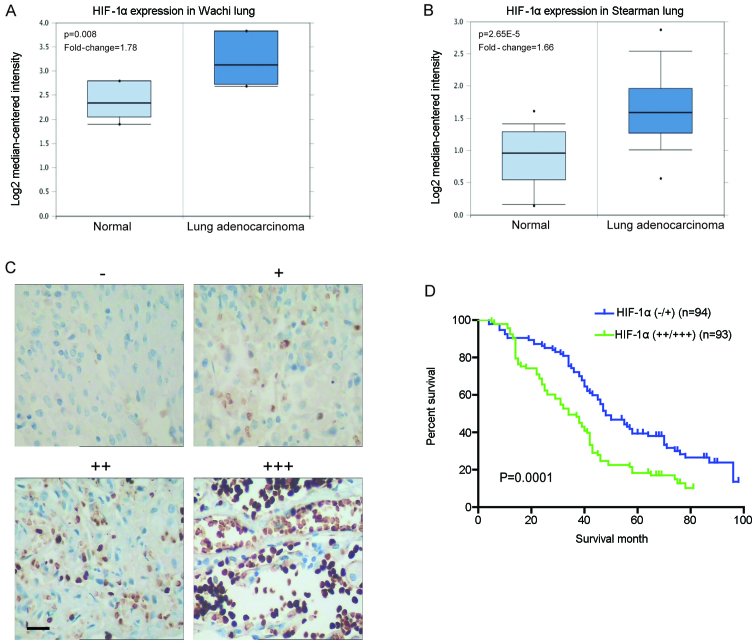
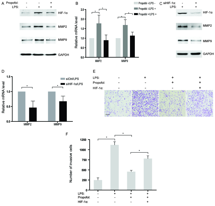
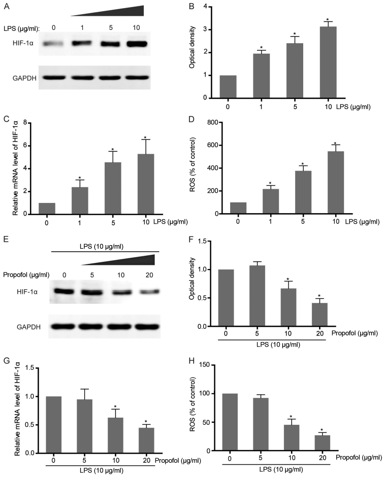
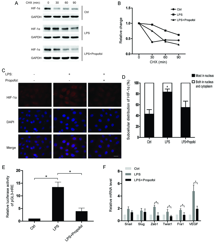
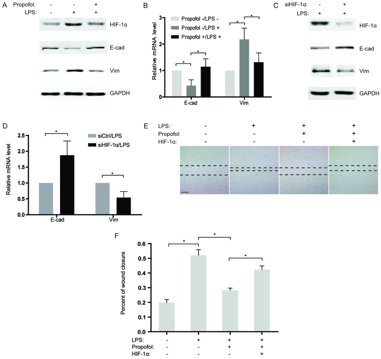
Tumor hypoxia has been recognized as a characteristic of the tumor microenvironment and promotes metastasis in a variety of types of cancer. However, in lung cancer, the role of hypoxia-inducible factor 1α (HIF-1α) in modulating the cellular response to the inflammation-related microenvironment remains unclear. In the present study, enhanced expression of HIF-1α accompanied by an increased ROS level was observed in lipopolysaccharide (LPS)-stimulated non-small cell lung cancer (NSCLC) cells. In addition, propofol, a general anesthetic, was found to significantly reduce the LPS-induced upregulation of HIF-1α and ROS in a dose-dependent manner. Further study showed that propofol may antagonize the role of LPS in activating HIF-1α through attenuating the protein stability and nuclear localization of HIF-1α. Moreover, knockdown of HIF-1α attenuated expression of mesenchymal marker, vimentin, but promoted the expression of epidermal marker, E-cadherin, in the LPS-treated NSCLC cells. Notably, LPS-induced epithelial-to-mesenchymal transition (EMT) was notably suppressed by propofol treatment. Consistently, a wound healing assay revealed that propofol abrogated LPS-stimulated migration of NSCLC cells while overexpression of HIF-1α reversed the effects of propofol. Similarly, we investigated the influence of propofol on the invasive capability of NSCLC cells. Western blot and RT-PCR analyses indicated that both knockdown of HIF-1α and treatment of propofol attenuated the LPS-activated expression of MMP2 and MMP9 which are necessary for tumor invasion. However, results from the Transwell assay confirmed that propofol also suppressed cell invasion by decreasing HIF-1α expression in the LPS-treated NSCLC cells. Analysis of clinical specimens demonstrated abnormal expression of HIF-1α in NSCLC tissues and a poor prognosis in patients with elevated HIF-1α expression. Thus, the present study suggests a potential strategy for NSCLC by targeting HIF-1α.
肿瘤缺氧已被认为是肿瘤微环境的一个特征,并在多种类型的癌症中促进转移。然而,在肺癌中,缺氧诱导因子1α(HIF-1α)在调节细胞对炎症相关微环境的反应中的作用仍不清楚。在本研究中,在脂多糖(LPS)刺激的非小细胞肺癌(NSCLC)细胞中观察到HIF-1α表达增强,同时活性氧(ROS)水平升高。此外,发现一种全身麻醉剂丙泊酚以剂量依赖性方式显著降低LPS诱导的HIF-1α和ROS上调。进一步研究表明,丙泊酚可能通过减弱HIF-1α的蛋白质稳定性和核定位来拮抗LPS激活HIF-1α的作用。此外,在LPS处理的NSCLC细胞中,敲低HIF-1α可减弱间充质标志物波形蛋白的表达,但促进表皮标志物E-钙黏蛋白的表达。值得注意的是,丙泊酚处理显著抑制了LPS诱导的上皮-间质转化(EMT)。同样,伤口愈合试验表明,丙泊酚消除了LPS刺激的NSCLC细胞迁移,而HIF-1α的过表达逆转了丙泊酚的作用。类似地,我们研究了丙泊酚对NSCLC细胞侵袭能力的影响。蛋白质印迹和逆转录-聚合酶链反应(RT-PCR)分析表明,敲低HIF-1α和丙泊酚处理均减弱了LPS激活的基质金属蛋白酶2(MMP2)和基质金属蛋白酶9(MMP9)的表达,这两者是肿瘤侵袭所必需的。然而,Transwell试验结果证实,丙泊酚还通过降低LPS处理的NSCLC细胞中HIF-1α的表达来抑制细胞侵袭。临床标本分析表明,NSCLC组织中HIF-1α表达异常,HIF-1α表达升高的患者预后较差。因此,本研究提出了一种通过靶向HIF-1α治疗NSCLC的潜在策略。