Department of Diabetes, Endocrinology and Nutrition, Graduate School of Medicine, Kyoto University, Kyoto, Japan.
Department of Genetics, Research Institute of Environmental Medicine, Nagoya University, Nagoya, Japan.
J Diabetes Investig. 2018 Jan;9(1):25-32. doi: 10.1111/jdi.12681. Epub 2017 May 29.
AIMS/INTRODUCTION: Glucagon-like peptide-1 (GLP-1) secreted from enteroendocrine L cells is an incretin that potentiates insulin secretion and is already applied in therapies for type 2 diabetes. However, detailed examination of L cells throughout the gastrointestinal tract remains unclear, because of difficulties in purifying scattered L cells from other cells. In the present study, we identified characteristics of L cells of the upper small intestine (UI), the lower small intestine (LI) and the colon using glucagon-green fluorescent protein-expressing mice that express GFP driven by the proglucagon promoter.
The localization and density of primary L cells were evaluated by anti-green fluorescent protein antibody reactivity. GLP-1 content, messenger ribonucleic acid (mRNA) expression levels and secretion in purified L cells were measured.
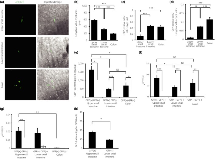
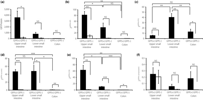
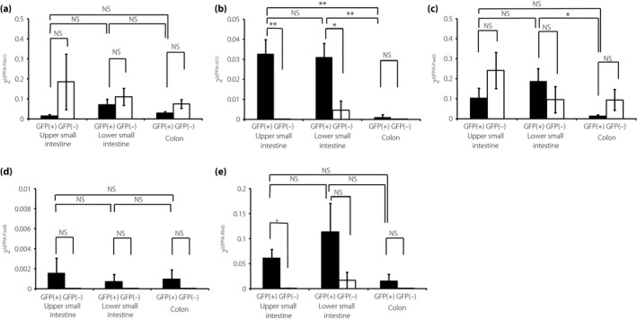
The number of L cells significantly increased toward the colon. In contrast, the GLP-1 content and secretion from L cells were higher in the UI than in the LI and colon. L cells from the UI and LI expressed notably high mRNA levels of the transcription factor, islet 1. The mRNA expression levels of peptide YY in L cells were higher in the LI than in the UI and colon. The mRNA expression levels of gastric inhibitory polypeptide in L cells from the UI were significantly higher compared with those from the LI and colon.
L cells show different numbers and characteristics throughout the gut, and they express different mRNA levels of transcription factors and gastrointestinal hormones. These results contribute to the therapeutic application of promoting GLP-1 release from L cells for the treatment of type 2 diabetes.
目的/引言:肠内分泌 L 细胞分泌的胰高血糖素样肽-1(GLP-1)是一种肠促胰岛素,可增强胰岛素分泌,已应用于 2 型糖尿病的治疗。然而,由于从其他细胞中纯化分散的 L 细胞存在困难,因此对整个胃肠道 L 细胞的详细检查仍不清楚。在本研究中,我们使用表达由前胰高血糖素启动子驱动 GFP 的胰高血糖素-绿色荧光蛋白表达小鼠,鉴定了上小肠(UI)、下小肠(LI)和结肠的 L 细胞的特征。
通过抗绿色荧光蛋白抗体反应性评估初级 L 细胞的定位和密度。测量纯化 L 细胞中的 GLP-1 含量、信使核糖核酸(mRNA)表达水平和分泌。
L 细胞的数量向结肠显著增加。相比之下,L 细胞的 GLP-1 含量和分泌在上小肠比在 LI 和结肠更高。UI 和 LI 的 L 细胞表达了明显高的转录因子胰岛 1 的 mRNA 水平。LI 中的 L 细胞中肽 YY 的 mRNA 表达水平高于 UI 和结肠。UI 中的 L 细胞中胃抑制肽的 mRNA 表达水平明显高于 LI 和结肠。
L 细胞在整个肠道中具有不同的数量和特征,并且它们表达不同的转录因子和胃肠激素的 mRNA 水平。这些结果有助于促进 L 细胞释放 GLP-1 用于治疗 2 型糖尿病的治疗的应用。