• 文献检索
  • 文档翻译
  • 深度研究
  • 学术资讯
  • Suppr Zotero 插件Zotero 插件
  • 邀请有礼
  • 套餐&价格
  • 历史记录
应用&插件
Suppr Zotero 插件Zotero 插件浏览器插件Mac 客户端Windows 客户端微信小程序
定价
高级版会员购买积分包购买API积分包
服务
文献检索文档翻译深度研究API 文档MCP 服务
关于我们
关于 Suppr公司介绍联系我们用户协议隐私条款
关注我们

Suppr 超能文献

核心技术专利:CN118964589B侵权必究
粤ICP备2023148730 号-1Suppr @ 2026

文献检索

告别复杂PubMed语法,用中文像聊天一样搜索,搜遍4000万医学文献。AI智能推荐,让科研检索更轻松。

立即免费搜索

文件翻译

保留排版,准确专业,支持PDF/Word/PPT等文件格式,支持 12+语言互译。

免费翻译文档

深度研究

AI帮你快速写综述,25分钟生成高质量综述,智能提取关键信息,辅助科研写作。

立即免费体验

使用锥形束计算机断层扫描对颞下颌关节紊乱患者和正常对照者的下颌髁突进行空间分析。

Spatial analysis of mandibular condyles in patients with temporomandibular disorders and normal controls using cone beam computed tomography.

作者信息

Al-Rawi Natheer Hashim, Uthman Asmaa Tahseen, Sodeify Sahar M

机构信息

Department Oral Health Sciences, College of Dental Medicine, University of Sharjah, Sharjah, UAE.

Department of Oral Medicine and Radiology, Gulf Medical University, Ajman, UAE.

出版信息

Eur J Dent. 2017 Jan-Mar;11(1):99-105. doi: 10.4103/ejd.ejd_202_16.

DOI:10.4103/ejd.ejd_202_16
PMID:28435374
原文链接:https://pmc.ncbi.nlm.nih.gov/articles/PMC5379844/
Abstract

OBJECTIVES

The aim of the study is to investigate the condylar position and its relation to articular eminence and axial condylar angle in temporomandibular joint disorder (TMD) patients and in normal controls using cone beam computed tomography (CBCT).

MATERIALS AND METHODS

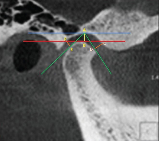
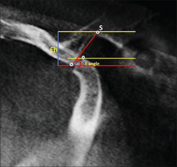
CBCT temporomandibular joint (TMJ) images of 70 participants (38 males and 32 females, mean age 26.4 years) were analyzed. They were divided into control group (including 35 subjects) and study group (including 35 subjects). Linear measurements of joint space and condyle determined the condylar position of each TMJ. Articular eminence height and inclination were also measured with axial condylar angle to determine its relation to condylar position. Independent and paired sample -test was applied to compare between the groups and TMJ sides of the same group at significance level of 0.05.

RESULTS

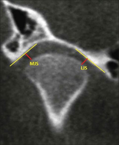
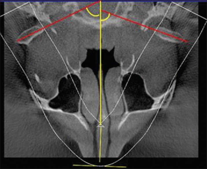
Statistical significant differences were found between males and females of both groups regarding superior joint space (SJS), lateral joint space, A-P, and M-L condyle distance ( < 0.05). SJS, medial joint space (MJS), and eminence angle were greater ( < 0.01) in male's joints with TMD with flatter axial condylar angle ( < 0.05), when compared with normal TMJ counterpart. Females TMJs showed significantly higher values of MJS of affected side when compared with normal counterpart with flatter axial condylar angle ( < 0.05).

CONCLUSION

Superior and MJS parameters were the ones that showed significant differences between affected and nonaffected joints. The mean axial condylar angle was smaller in joints with abnormal TMJ. This indicates that the condyles of the affected joints may rotate inward.

摘要

目的

本研究旨在使用锥形束计算机断层扫描(CBCT),调查颞下颌关节紊乱病(TMD)患者和正常对照者的髁突位置及其与关节结节和髁突轴角的关系。

材料与方法

分析了70名参与者(38名男性和32名女性,平均年龄26.4岁)的CBCT颞下颌关节(TMJ)图像。他们被分为对照组(包括35名受试者)和研究组(包括35名受试者)。通过对关节间隙和髁突的线性测量确定每个TMJ的髁突位置。还测量了关节结节高度和倾斜度以及髁突轴角,以确定其与髁突位置的关系。采用独立样本和配对样本t检验,在0.05的显著性水平下比较两组之间以及同一组TMJ两侧之间的差异。

结果

两组的男性和女性在关节上间隙(SJS)、外侧关节间隙、前后和内外髁突距离方面均存在统计学显著差异(P<0.05)。与正常TMJ相比,患有TMD的男性关节的SJS、内侧关节间隙(MJS)和关节结节角更大(P<0.01),髁突轴角更平坦(P<0.05)。与正常对应侧相比,女性患侧TMJ的MJS值显著更高,髁突轴角更平坦(P<0.05)。

结论

上间隙和MJS参数在患侧和未患侧关节之间显示出显著差异。TMJ异常的关节的平均髁突轴角较小。这表明患侧关节的髁突可能向内旋转。

https://cdn.ncbi.nlm.nih.gov/pmc/blobs/47b2/5379844/2b831b80c931/EJD-11-99-g004.jpg
https://cdn.ncbi.nlm.nih.gov/pmc/blobs/47b2/5379844/d3461e05af3a/EJD-11-99-g001.jpg
https://cdn.ncbi.nlm.nih.gov/pmc/blobs/47b2/5379844/0641bab9eaef/EJD-11-99-g002.jpg
https://cdn.ncbi.nlm.nih.gov/pmc/blobs/47b2/5379844/631376f2bb36/EJD-11-99-g003.jpg
https://cdn.ncbi.nlm.nih.gov/pmc/blobs/47b2/5379844/2b831b80c931/EJD-11-99-g004.jpg

相似文献

1
Spatial analysis of mandibular condyles in patients with temporomandibular disorders and normal controls using cone beam computed tomography.使用锥形束计算机断层扫描对颞下颌关节紊乱患者和正常对照者的下颌髁突进行空间分析。
Eur J Dent. 2017 Jan-Mar;11(1):99-105. doi: 10.4103/ejd.ejd_202_16.
2
Age and Gender-related Morphometric Assessment and Degenerative Changes of Temporomandibular Joint in Symptomatic Subjects and Controls using Cone Beam Computed Tomography (CBCT): A Comparative Analysis.使用锥形束计算机断层扫描 (CBCT) 对有症状的受试者和对照者进行年龄和性别相关的形态计量评估和颞下颌关节退行性改变:比较分析。
Curr Med Imaging. 2024;20:1-12. doi: 10.2174/0115734056248617231002110417.
3
[Cone-beam CT evaluation of temporomandibular joint in skeletal class Ⅱ female adolescents with different vertical patterns].[锥形束CT对不同垂直骨面型Ⅱ类女性青少年颞下颌关节的评估]
Beijing Da Xue Xue Bao Yi Xue Ban. 2020 Dec 29;53(1):109-119. doi: 10.19723/j.issn.1671-167X.2021.01.017.
4
Quantitative and qualitative condylar changes following stabilization splint therapy in patients with temporomandibular joint disorders with and without skeletal lateral mandibular asymmetry: a cone beam computed tomographic study.髁突定量和定性变化在伴有和不伴有下颌骨横向不对称的颞下颌关节紊乱患者的稳定夹板治疗后的变化:锥形束 CT 研究。
BMC Oral Health. 2024 Mar 21;24(1):363. doi: 10.1186/s12903-024-04119-7.
5
Positional, morphologic, and volumetric differences in TMJ in unilateral posterior crossbites and controls: A retrospective CBCT study.单侧后牙反合与对照组颞下颌关节的位置、形态和容积差异:一项回顾性CBCT研究
Int Orthod. 2024 Sep;22(3):100889. doi: 10.1016/j.ortho.2024.100889. Epub 2024 Jun 3.
6
Comparative cone-beam computed tomography evaluation of the osseous morphology of the temporomandibular joint in temporomandibular dysfunction patients and asymptomatic individuals.颞下颌关节紊乱病患者与无症状个体颞下颌关节骨形态的锥束计算机断层扫描对比评估
Oral Radiol. 2018 Jan;34(1):31-39. doi: 10.1007/s11282-017-0279-7. Epub 2017 Mar 16.
7
Comparative assessment of condylar position in patients with temporomandibular disorder (TMD) and asymptomatic patients using cone-beam computed tomography.使用锥形束计算机断层扫描对颞下颌关节紊乱病(TMD)患者和无症状患者的髁突位置进行比较评估。
Dent Med Probl. 2019 Jan-Mar;56(1):81-87. doi: 10.17219/dmp/102946.
8
Comparison of temporomandibular joints in relation to ages and vertical facial types in skeletal class II female patients: a multiple-cross-sectional study.比较不同年龄和垂直面型的骨性 II 类女性患者颞下颌关节:一项多横断面研究。
BMC Oral Health. 2024 Apr 17;24(1):467. doi: 10.1186/s12903-024-04219-4.
9
Assessing joint space and condylar position in the people with normal function of temporomandibular joint with cone-beam computed tomography.使用锥形束计算机断层扫描评估颞下颌关节功能正常人群的关节间隙和髁突位置。
Dent Res J (Isfahan). 2012 Sep;9(5):607-12. doi: 10.4103/1735-3327.104881.
10
Evaluation of condylar positions in patients with temporomandibular disorders: A cone-beam computed tomographic study.颞下颌关节紊乱病患者髁突位置的评估:一项锥形束计算机断层扫描研究。
Imaging Sci Dent. 2016 Jun;46(2):127-31. doi: 10.5624/isd.2016.46.2.127. Epub 2016 Jun 23.

引用本文的文献

1
Optimization of the size and location of the FOVs for CBCT capture of the TMJ.锥形束计算机断层扫描(CBCT)采集颞下颌关节(TMJ)时视野(FOV)大小和位置的优化。
BMC Oral Health. 2025 May 10;25(1):709. doi: 10.1186/s12903-025-06081-4.
2
Volumetric Comparison of the Temporomandibular Joint Space in Skeletal Class I and II Patients Using Cone-Beam Computed Tomography: A Cross-Sectional Study.使用锥形束计算机断层扫描对骨骼Ⅰ类和Ⅱ类患者颞下颌关节间隙进行容积比较:一项横断面研究。
Cureus. 2024 Dec 12;16(12):e75597. doi: 10.7759/cureus.75597. eCollection 2024 Dec.
3
Modifications of the articular eminence inclination in association with the loss of occlusal support in the molar area. A cone-beam computed tomography study.

本文引用的文献

1
Cone-beam tomography assessment of the condylar position in asymptomatic and symptomatic young individuals.锥形束断层扫描对无症状和有症状的年轻个体髁突位置的评估。
J Prosthet Dent. 2015 Sep;114(3):420-5. doi: 10.1016/j.prosdent.2015.04.006. Epub 2015 Jun 2.
2
Association between mandibular condylar position and clinical dysfunction index.下颌髁突位置与临床功能障碍指数之间的关联。
J Craniomaxillofac Surg. 2015 May;43(4):432-6. doi: 10.1016/j.jcms.2015.01.005. Epub 2015 Jan 24.
3
Clinical and radiological findings of a bilateral coronoid hyperplasia case.
髁突前斜面倾斜度的改变与磨牙后区咬合支持丧失的相关性:锥形束 CT 研究。
Rom J Morphol Embryol. 2024 Apr-Jun;65(2):297-307. doi: 10.47162/RJME.65.2.17.
4
Assessment of condylar anatomy and degenerative changes in temporomandibular joint disorders - A scoping review.颞下颌关节紊乱病中髁突解剖结构及退行性改变的评估——一项范围综述
J Oral Biol Craniofac Res. 2023 Nov-Dec;13(6):764-780. doi: 10.1016/j.jobcr.2023.10.004. Epub 2023 Nov 6.
5
Condylar Positional Changes in Skeletal Class II and Class III Malocclusions after Bimaxillary Orthognathic Surgery.双颌正颌外科手术后骨性II类和III类错牙合畸形髁突位置的变化
J Pers Med. 2023 Oct 27;13(11):1544. doi: 10.3390/jpm13111544.
6
Assessment of bony changes in temporomandibular joint in patients using cone beam computed tomography - a cross sectional study.利用锥形束 CT 评估颞下颌关节的骨改变 - 一项横断面研究。
Head Face Med. 2023 Oct 28;19(1):47. doi: 10.1186/s13005-023-00392-z.
7
Dimensional and positional temporomandibular joint osseous characteristics in normodivergent facial patterns with and without temporomandibular disorders.正常颅面型伴或不伴颞下颌关节紊乱患者的颞下颌关节骨性特征的三维位置分析。
Clin Oral Investig. 2023 Sep;27(9):5011-5020. doi: 10.1007/s00784-023-05120-0. Epub 2023 Jun 26.
8
Positional Features of the Mandibular Condyle in Patients with Facial Asymmetry.面部不对称患者下颌髁突的位置特征
Diagnostics (Basel). 2023 Mar 8;13(6):1034. doi: 10.3390/diagnostics13061034.
9
Cone beam computed tomography in the assessment of TMJ deformity in children with JIA: repeatability of a novel scoring system.锥形束 CT 在儿童 JIA 伴 TMJ 畸形评估中的应用:新型评分系统的可重复性。
BMC Oral Health. 2023 Jan 10;23(1):12. doi: 10.1186/s12903-022-02701-5.
10
Three-Dimensional Assessment of the Temporomandibular Joint Changes Following Reversed Twin Block Therapy of Patients With Skeletal Class III Malocclusion in Conjunction With the Photobiomodulation Therapy: A Randomized Controlled Clinical Trial.三维评估骨骼Ⅲ类错牙合患者采用反向 Twin Block 矫治联合光生物调节疗法后的颞下颌关节变化:一项随机对照临床试验
Cureus. 2022 Jun 13;14(6):e25897. doi: 10.7759/cureus.25897. eCollection 2022 Jun.
一例双侧冠状突增生病例的临床及影像学表现
Eur J Dent. 2015 Jan-Mar;9(1):149-152. doi: 10.4103/1305-7456.149665.
4
Three-dimensional imaging techniques: A literature review.三维成像技术:文献综述。
Eur J Dent. 2014 Jan;8(1):132-140. doi: 10.4103/1305-7456.126269.
5
Assessing joint space and condylar position in the people with normal function of temporomandibular joint with cone-beam computed tomography.使用锥形束计算机断层扫描评估颞下颌关节功能正常人群的关节间隙和髁突位置。
Dent Res J (Isfahan). 2012 Sep;9(5):607-12. doi: 10.4103/1735-3327.104881.
6
Osteoarthritic changes and condylar positioning of the temporomandibular joint in Korean children and adolescents.韩国儿童和青少年颞下颌关节的骨关节炎变化及髁突位置
Imaging Sci Dent. 2012 Sep;42(3):169-74. doi: 10.5624/isd.2012.42.3.169. Epub 2012 Sep 21.
7
Relationship between the thickness of the roof of glenoid fossa, condyle morphology and remaining teeth in asymptomatic European patients based on cone beam CT data sets.基于锥形束 CT 数据集的无症状欧洲患者关节窝顶厚度、髁突形态与余留牙的关系。
Dentomaxillofac Radiol. 2013;42(3):90929410. doi: 10.1259/dmfr/90929410. Epub 2012 Sep 20.
8
The Research Diagnostic Criteria for Temporomandibular Disorders. VI: future directions.颞下颌关节紊乱病的研究诊断标准。第六章:未来方向。
J Orofac Pain. 2010 Winter;24(1):79-88.
9
Assessment of optimal condylar position with limited cone-beam computed tomography.使用有限锥形束计算机断层扫描评估髁突最佳位置
Am J Orthod Dentofacial Orthop. 2009 Apr;135(4):495-501. doi: 10.1016/j.ajodo.2007.05.021.
10
Asymmetry of the articular eminence in dentate and partially edentulous populations.有牙列和部分无牙列人群中关节结节的不对称性。
Cranio. 2006 Apr;24(2):85-94. doi: 10.1179/crn.2006.014.