• 文献检索
  • 文档翻译
  • 深度研究
  • 学术资讯
  • Suppr Zotero 插件Zotero 插件
  • 邀请有礼
  • 套餐&价格
  • 历史记录
应用&插件
Suppr Zotero 插件Zotero 插件浏览器插件Mac 客户端Windows 客户端微信小程序
定价
高级版会员购买积分包购买API积分包
服务
文献检索文档翻译深度研究API 文档MCP 服务
关于我们
关于 Suppr公司介绍联系我们用户协议隐私条款
关注我们

Suppr 超能文献

核心技术专利:CN118964589B侵权必究
粤ICP备2023148730 号-1Suppr @ 2026

文献检索

告别复杂PubMed语法,用中文像聊天一样搜索,搜遍4000万医学文献。AI智能推荐,让科研检索更轻松。

立即免费搜索

文件翻译

保留排版,准确专业,支持PDF/Word/PPT等文件格式,支持 12+语言互译。

免费翻译文档

深度研究

AI帮你快速写综述,25分钟生成高质量综述,智能提取关键信息,辅助科研写作。

立即免费体验

膝关节半月板挤出:仅3毫米的挤出就应被视为显著吗?通过MRI和关节镜检查进行的评估。

Meniscal Extrusion in the Knee: Should only 3 mm Extrusion be Considered Significant? An Assessment by MRI and Arthroscopy.

作者信息

Muzaffar N, Kirmani O, Ahsan M, Ahmad S

机构信息

Hospital for Bone and Joint Surgery, Srinagar, India.

Department of Radiodiagnosis, Government Medical College, Srinagar, India.

出版信息

Malays Orthop J. 2015 Jul;9(2):17-20. doi: 10.5704/MOJ.1507.013.

DOI:10.5704/MOJ.1507.013
PMID:28435604
原文链接:https://pmc.ncbi.nlm.nih.gov/articles/PMC5333659/
Abstract

AIM

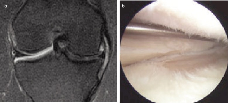
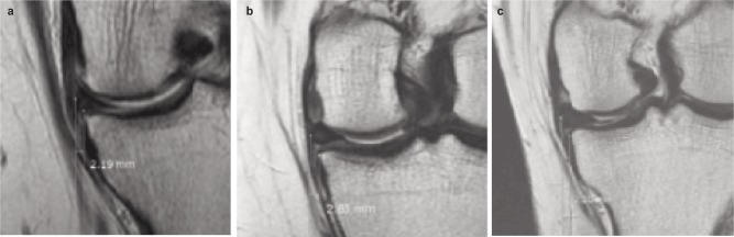
The aim of this study was to assess whether significant meniscal extrusion of more than 3 mm or of even lesser degrees of extrusion could be considered significant. We also aimed to determine the morphology of tears that are most likely to be associated with significant extrusion. Study design and material. The study was done retrospectively on a group of 202 patients (157 males and 45 females) who had been seen in our hospital between 2007 and 2011 with meniscal tears (in one knee only) diagnosed by MRI and confirmed on arthroscopy. Extrusion of 3 mm or more (usually considered significant) was seen in 102 cases and less than 3 mm in 100. Extrusion was measured on the coronal MR images rather than on saggital images because of ease and reproducibility. The tears were confirmed by arthroscopy and correlated with the extent of extrusion on MRI.

RESULTS

Out of the total of 202 cases, 102 cases (50.5%) had extrusion of 3 mm or more on MRI. Of these, the medial meniscal posterior horn tears accounted for 63 cases (64.26%), 21 cases were medial meniscal body tears (21.42%), five medial meniscal root tears (5.1%), nine lateral meniscal body tears (9.18%) and four lateral meniscal posterior horn tears(4.08%). Forty-four cases had extrusion of 3-4 mm, 26 had extrusion of 4-5mm, 17 cases had extrusion of 5-6mm, ten had extrusion of 6-7mm and five had extrusion of 7 mm or more. One hundred cases fell in the < 3mm extrusion category, of which 80 (39.6%) were in the 2-3 mm extrusion group and 20 (9.9%) in the 1-2 mm extrusion group. They comprised of 61 cases of medial meniscal posterior horn tears, 23 cases of medial meniscal body tears, six medial meniscal root tears, eight lateral meniscal body tears and two lateral meniscal posterior horn tears. The highest proportion of meniscal tears was seen in the 2-3 mm category comprising nearly 40% of the entire study group. The majority of tears were medial meniscal posterior horn tears.

CONCLUSION

Menisci that extruded 2-3 mm from the tibial margin formed a major proportion of menisci treated for tears by repair or menisectomy. We should consider extrusion of more than 2mm as significant. Most tears had extrusion of 2-4 mm.

摘要

目的

本研究旨在评估半月板挤出超过3mm或甚至更小程度的挤出是否可被视为有意义。我们还旨在确定最有可能与明显挤出相关的撕裂形态。研究设计与材料。本研究对2007年至2011年期间在我院就诊的一组202例患者(157例男性和45例女性)进行回顾性研究,这些患者经MRI诊断为半月板撕裂(仅累及一侧膝关节)并经关节镜检查证实。102例患者半月板挤出3mm或更多(通常认为有意义),100例患者挤出小于3mm。由于简便性和可重复性,在冠状面MR图像上测量挤出情况,而非矢状面图像。撕裂情况经关节镜检查证实,并与MRI上的挤出程度相关联。

结果

在总共202例病例中,102例(50.5%)在MRI上挤出3mm或更多。其中,内侧半月板后角撕裂占63例(64.26%),内侧半月板体部撕裂21例(21.42%),内侧半月板根部撕裂5例(5.1%),外侧半月板体部撕裂9例(9.18%),外侧半月板后角撕裂4例(4.08%)。44例挤出3 - 4mm,26例挤出4 - 5mm,17例挤出5 - 6mm,1个例挤出6 - 7mm,5例挤出7mm或更多。100例属于挤出<3mm类别,其中80例(39.6%)在2 - 3mm挤出组,20例(9.

https://cdn.ncbi.nlm.nih.gov/pmc/blobs/4158/5333659/838cf41fd15c/moj-9-017-f3.jpg
https://cdn.ncbi.nlm.nih.gov/pmc/blobs/4158/5333659/076079093dd5/moj-9-017-f1.jpg
https://cdn.ncbi.nlm.nih.gov/pmc/blobs/4158/5333659/ef77b22e5b45/moj-9-017-f2.jpg
https://cdn.ncbi.nlm.nih.gov/pmc/blobs/4158/5333659/838cf41fd15c/moj-9-017-f3.jpg
https://cdn.ncbi.nlm.nih.gov/pmc/blobs/4158/5333659/076079093dd5/moj-9-017-f1.jpg
https://cdn.ncbi.nlm.nih.gov/pmc/blobs/4158/5333659/ef77b22e5b45/moj-9-017-f2.jpg
https://cdn.ncbi.nlm.nih.gov/pmc/blobs/4158/5333659/838cf41fd15c/moj-9-017-f3.jpg

相似文献

1
Meniscal Extrusion in the Knee: Should only 3 mm Extrusion be Considered Significant? An Assessment by MRI and Arthroscopy.膝关节半月板挤出:仅3毫米的挤出就应被视为显著吗?通过MRI和关节镜检查进行的评估。
Malays Orthop J. 2015 Jul;9(2):17-20. doi: 10.5704/MOJ.1507.013.
2
Medial extrusion of the posterior segment of medial meniscus is a sensitive sign for posterior horn tears.内侧半月板后段的内侧挤压是后角撕裂的敏感征象。
Knee. 2014 Jan;21(1):112-8. doi: 10.1016/j.knee.2013.02.003. Epub 2013 Apr 25.
3
MR findings of meniscal extrusion correlated with arthroscopy.半月板挤出的磁共振成像(MR)表现与关节镜检查结果相关。
J Magn Reson Imaging. 2008 Aug;28(2):466-70. doi: 10.1002/jmri.21460.
4
Medial meniscus extrusion on knee MRI: is extent associated with severity of degeneration or type of tear?膝关节MRI上内侧半月板挤出:其程度与退变严重程度或撕裂类型有关吗?
AJR Am J Roentgenol. 2004 Jul;183(1):17-23. doi: 10.2214/ajr.183.1.1830017.
5
Comparison of Medial and Lateral Meniscus Root Tears.内侧半月板根部撕裂与外侧半月板根部撕裂的比较
PLoS One. 2015 Oct 21;10(10):e0141021. doi: 10.1371/journal.pone.0141021. eCollection 2015.
6
Magnetic resonance imaging evidence of meniscal extrusion in medial meniscus posterior root tear.内侧半月板后根撕裂的半月板挤压的磁共振成像证据。
Arthroscopy. 2010 Dec;26(12):1602-6. doi: 10.1016/j.arthro.2010.05.004.
7
Posterior horn medial meniscal root tear: the prequel.后角内侧半月板根部撕裂:前传
Skeletal Radiol. 2014 Jun;43(6):775-80. doi: 10.1007/s00256-014-1837-2. Epub 2014 Feb 15.
8
Tear gap and severity of osteoarthritis are associated with meniscal extrusion in degenerative medial meniscus posterior root tears.半月板后根撕裂中,撕裂间隙和骨关节炎严重程度与半月板外突有关。
Orthop Traumatol Surg Res. 2019 Nov;105(7):1395-1399. doi: 10.1016/j.otsr.2019.09.015. Epub 2019 Sep 28.
9
[Diagnostic value of MRI for posterior root tear of medial and lateral meniscus].[MRI对内外侧半月板后根撕裂的诊断价值]
Zhongguo Gu Shang. 2018 Mar 25;31(3):263-266. doi: 10.3969/j.issn.1003-0034.2018.03.014.
10
MR diagnosis of meniscal tears of the knee: importance of high signal in the meniscus that extends to the surface.膝关节半月板撕裂的磁共振成像诊断:半月板内延伸至表面的高信号的重要性。
AJR Am J Roentgenol. 1993 Jul;161(1):101-7. doi: 10.2214/ajr.161.1.8517286.

引用本文的文献

1
Influence of preoperative medial meniscus extrusion and subchondral bone marrow edema on outcomes after medial opening wedge high tibial osteotomy.术前内侧半月板挤压和软骨下骨髓水肿对内侧开放楔形高位胫骨截骨术后疗效的影响。
J Orthop Surg Res. 2025 Apr 9;20(1):353. doi: 10.1186/s13018-025-05656-9.
2
Meniscal extrusion: Proposal for a novel qualitative classification.半月板挤压:一种新型定性分类的建议
J Exp Orthop. 2024 Dec 31;12(1):e70126. doi: 10.1002/jeo2.70126. eCollection 2025 Jan.
3
The Relationship Between Medial Meniscal Extrusion and Outcome Measures for Knee Osteoarthritis: A Systematic Review.

本文引用的文献

1
MR findings of meniscal extrusion correlated with arthroscopy.半月板挤出的磁共振成像(MR)表现与关节镜检查结果相关。
J Magn Reson Imaging. 2008 Aug;28(2):466-70. doi: 10.1002/jmri.21460.
2
Relationship of meniscal damage, meniscal extrusion, malalignment, and joint laxity to subsequent cartilage loss in osteoarthritic knees.骨关节炎膝关节中半月板损伤、半月板挤出、力线不正及关节松弛与后续软骨丢失的关系。
Arthritis Rheum. 2008 Jun;58(6):1716-26. doi: 10.1002/art.23462.
3
Meniscal extrusion in young athletes: associated knee joint abnormalities.
内侧半月板挤压与膝关节骨关节炎结局指标之间的关系:一项系统评价
Orthop J Sports Med. 2024 Aug 9;12(8):23259671241248457. doi: 10.1177/23259671241248457. eCollection 2024 Aug.
4
Efficacy of a single ultrasound-guided injection of high molecular weight hyaluronic acid combined with collagen tripeptide in patients with knee osteoarthritis and chondrocalcinosis.单次超声引导下注射高分子量透明质酸联合胶原三肽治疗膝骨关节炎合并软骨钙质沉着症的疗效
Front Med (Lausanne). 2024 Jul 19;11:1437160. doi: 10.3389/fmed.2024.1437160. eCollection 2024.
5
Seven tesla knee MRI T2*-mapping detects intrasubstance meniscus degeneration in patients with posterior root tears.7特斯拉膝关节MRI T2*成像可检测后交叉韧带撕裂患者半月板实质内的退变。
Radiol Adv. 2024 Mar 19;1(1):umae005. doi: 10.1093/radadv/umae005. eCollection 2024 May.
6
Measuring Technique for Meniscal Extrusion Using Ultrasound in the Setting of Posterior Medial Meniscal Root Tears.后内侧半月板根部撕裂情况下使用超声测量半月板挤出的技术
Arthrosc Tech. 2024 Jan 30;13(4):102916. doi: 10.1016/j.eats.2024.102916. eCollection 2024 Apr.
7
Meniscal extrusion: risk factors and diagnostic tools to predict early osteoarthritis.半月板挤出:预测早期骨关节炎的危险因素和诊断工具。
Orthop Rev (Pavia). 2023 May 13;15:74881. doi: 10.52965/001c.74881. eCollection 2023.
8
Clinical significance and management of meniscal extrusion in different knee pathologies: a comprehensive review of the literature and treatment algorithm.不同膝关节病变中半月板挤压的临床意义及处理:文献综述与治疗方案
Knee Surg Relat Res. 2022 Jul 18;34(1):35. doi: 10.1186/s43019-022-00163-1.
9
Meniscal Extrusion: A Narrative Review.半月板挤出:一篇叙述性综述。
Orthop J Sports Med. 2021 Nov 3;9(11):23259671211043797. doi: 10.1177/23259671211043797. eCollection 2021 Nov.
10
Sonographic evaluation of the degree of medial meniscal extrusion during Thessaly test in healthy knees.在健康膝关节 Thessaly 试验中评估内侧半月板外突程度的超声评估。
Chiropr Man Therap. 2021 Aug 17;29(1):31. doi: 10.1186/s12998-021-00390-5.
年轻运动员的半月板挤出:相关的膝关节异常情况。
AJR Am J Roentgenol. 2006 Mar;186(3):791-4. doi: 10.2214/AJR.04.1181.
4
The role of meniscal root pathology and radial meniscal tear in medial meniscal extrusion.半月板根部病变和半月板放射状撕裂在半月板内侧挤压中的作用。
Skeletal Radiol. 2004 Oct;33(10):569-74. doi: 10.1007/s00256-004-0761-2. Epub 2004 Aug 14.
5
Medial meniscus extrusion on knee MRI: is extent associated with severity of degeneration or type of tear?膝关节MRI上内侧半月板挤出:其程度与退变严重程度或撕裂类型有关吗?
AJR Am J Roentgenol. 2004 Jul;183(1):17-23. doi: 10.2214/ajr.183.1.1830017.
6
Radial displacement of the medial meniscus in varus osteoarthritis of the knee.膝关节内翻性骨关节炎中内侧半月板的径向移位。
Clin Orthop Relat Res. 2001 Jun(387):171-7. doi: 10.1097/00003086-200106000-00023.
7
Meniscal movement. An in-vivo study using dynamic MRI.半月板运动。一项使用动态磁共振成像的体内研究。
J Bone Joint Surg Br. 1999 Jan;81(1):37-41. doi: 10.1302/0301-620x.81b1.8928.
8
Meniscal position on routine MR imaging of the knee.膝关节常规磁共振成像时半月板的位置
Skeletal Radiol. 1997 Jul;26(7):424-7. doi: 10.1007/s002560050259.
9
Radial displacement of the medial meniscus and Fairbank's signs.内侧半月板的径向移位及费尔班克氏征
Clin Orthop Relat Res. 1997 Jun(339):163-73. doi: 10.1097/00003086-199706000-00022.
10
Extrusion of the medial meniscus.内侧半月板挤出。
Arthroscopy. 1991;7(3):297-300. doi: 10.1016/0749-8063(91)90131-g.