Karlsson Tobias E, Wellfelt Katrin, Olson Lars
Department of Neuroscience, Karolinska InstitutetStockholm, Sweden.
Front Mol Neurosci. 2017 Apr 11;10:94. doi: 10.3389/fnmol.2017.00094. eCollection 2017.
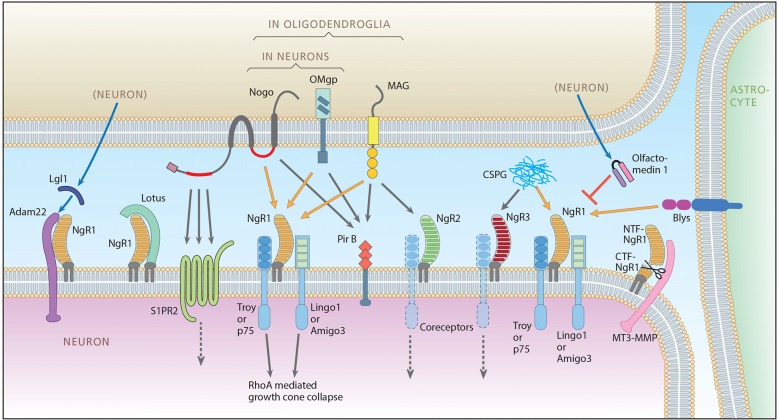
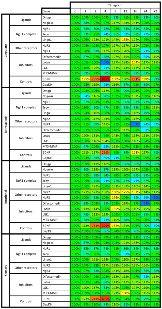
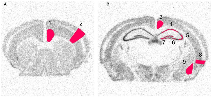
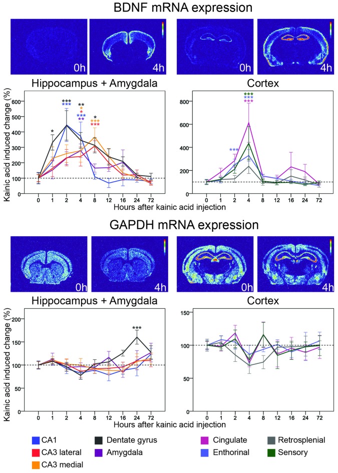
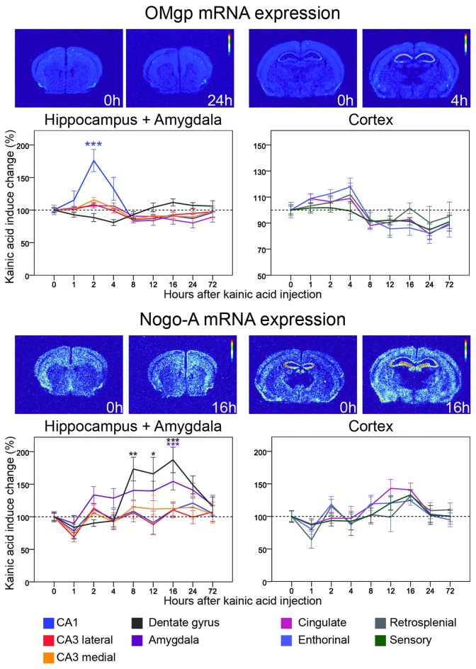
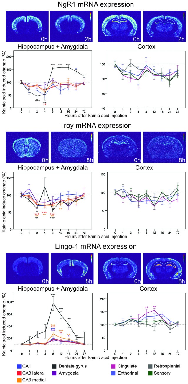
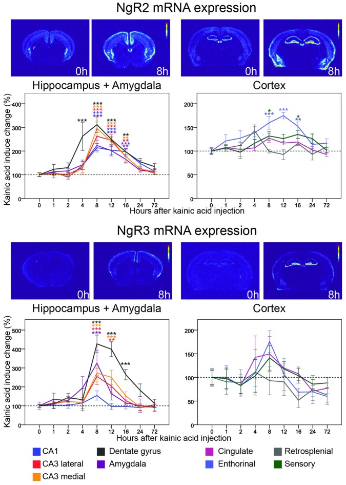
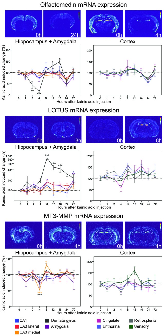
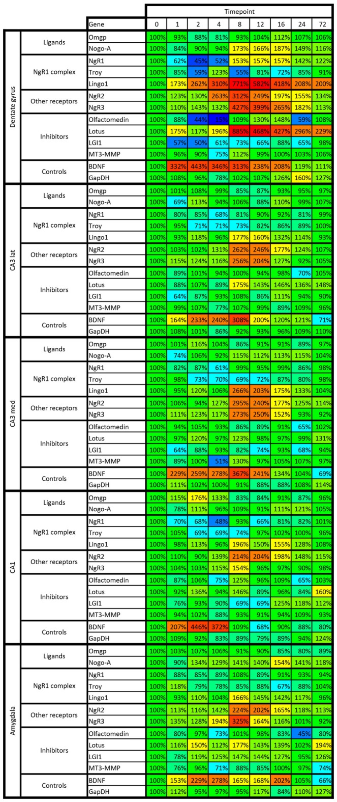
Inhibition of nerve growth and plasticity in the CNS is to a large part mediated by Nogo-like signaling, now encompassing a plethora of ligands, receptors, co-receptors and modulators. Here we describe the distribution and levels of mRNA encoding 11 key genes involved in Nogo-like signaling (Nogo-A, Oligodendrocyte-Myelin glycoprotein (OMgp), Nogo receptor 1 (NgR1), NgR2, NgR3, Lingo-1, TNF receptor orphan Y (Troy), Olfactomedin, Lateral olfactory tract usher substance (Lotus) and membrane-type matrix metalloproteinase-3 (MT3-MPP)), as well as BDNF and GAPDH. Expression was analyzed in nine different brain areas before, and at eight time points during the first 3 days after a strong neuroexcitatory stimulation, caused by one kainic acid injection. A temporo-spatial pattern of orderly transcriptional regulations emerges that strengthens the role of Nogo-signaling mechanisms for synaptic plasticity in synchrony with transcriptional increases of BDNF mRNA. For most Nogo-type signaling genes, the largest alterations of mRNA levels occur in the dentate gyrus, with marked alterations also in the CA1 region. Changes occurred somewhat later in several areas of the cerebral cortex. The detailed spatio-temporal pattern of mRNA presence and kainic acid-induced transcriptional response is gene-specific. We reveal that several different gene alterations combine to decrease (and later increase) Nogo-like signaling, as expected to allow structural plasticity responses. Other genes are altered in the opposite direction, suggesting that the system prepares in advance in order to rapidly restore balance. However, the fact that Lingo-1 shows a seemingly opposite, plasticity inhibiting response to kainic acid (strong increase of mRNA in the dentate gyrus), may instead suggest a plasticity-enhancing intracellular function of this presumed NgR1 co-receptor.
中枢神经系统中神经生长和可塑性的抑制在很大程度上是由类Nogo信号介导的,现在该信号包括大量的配体、受体、共受体和调节剂。在这里,我们描述了参与类Nogo信号传导的11个关键基因(Nogo-A、少突胶质细胞髓磷脂糖蛋白(OMgp)、Nogo受体1(NgR1)、NgR2、NgR3、Lingo-1、肿瘤坏死因子受体孤儿Y(Troy)、嗅觉介质、外侧嗅束引导物质(Lotus)和膜型基质金属蛋白酶-3(MT3-MMP))以及脑源性神经营养因子(BDNF)和甘油醛-3-磷酸脱氢酶(GAPDH)的mRNA分布和水平。在一次 kainic 酸注射引起的强烈神经兴奋刺激之前以及刺激后的前3天内的8个时间点,对九个不同脑区的表达进行了分析。出现了一种有序转录调控的时空模式,这加强了Nogo信号机制在与BDNF mRNA转录增加同步的突触可塑性中的作用。对于大多数Nogo型信号基因,mRNA水平的最大变化发生在齿状回,CA1区域也有明显变化。大脑皮层的几个区域的变化出现得稍晚一些。mRNA存在和 kainic 酸诱导的转录反应的详细时空模式是基因特异性的。我们发现,几种不同的基因改变共同作用以降低(随后增加)类Nogo信号,正如预期的那样,以允许结构可塑性反应。其他基因的改变方向相反,这表明该系统提前做好准备以便迅速恢复平衡。然而,Lingo-1对 kainic 酸表现出看似相反的、抑制可塑性的反应(齿状回中mRNA强烈增加)这一事实,可能反而表明这种假定的NgR1共受体具有增强可塑性的细胞内功能。