Gai Pengzhou, Sun Hongliang, Wang Guangda, Xu Qiang, Qi Xiaojun, Zhang Zuofu, Jiang Lei
Department of Orthopedics, Yantai Yuhuangding Hospital, Yantai, Shandong 264000, P.R. China.
Department of Pathology, Yantai Yuhuangding Hospital, Yantai, Shandong 264000, P.R. China.
Oncol Lett. 2017 Apr;13(4):2354-2358. doi: 10.3892/ol.2017.5674. Epub 2017 Feb 2.
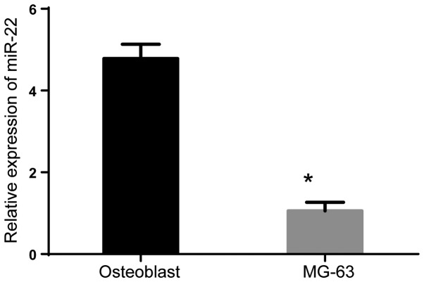
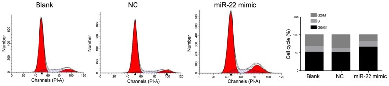
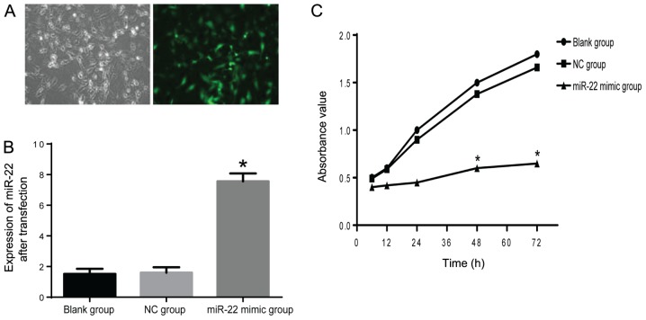
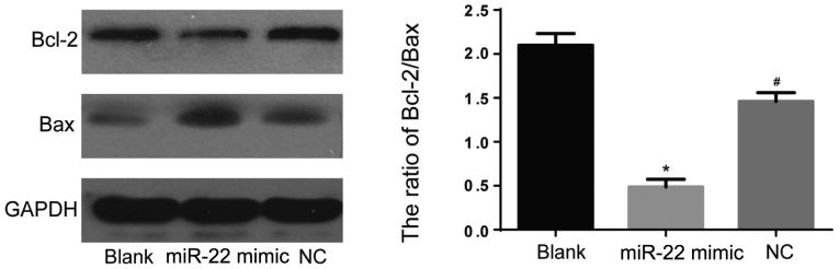
To study the effects of miR-22 on the proliferation and the apoptosis of osteosarcoma MG-63 cell line and to explore the potential molecular mechanism that miR-22 regulates this biological process. Quantitive real-time polymerase chain reaction (RT-qPCR) was performed to explore the miRNA level of miR-22. The MG-63 cell line was infected with miR-22 mimics for establishment of miR-22 overexpression. Non-infected cells were in blank group and cells infected with empty vector were served as negative control (NC group). MTT assay was conducted to measure cell viability. The cell cycle and apoptosis were explored using flow cytometry and the apoptosis-related markers were detected by western blotting. RT-qPCR results revealed that the miR-22 miRNA level in the MG-63 cells was significantly lower than that in osteoblasts (P<0.05). MTT assay showed that the MG-63 cells infected with miR-22 mimics exhibited markedly decreased proliferation ability compared with blank and empty vector (NC) groups. Next, we found that overexpression of miR-22 remarkably increased the apoptosis of the MG-63 cells, evidenced from the flow cytometry results and elevated Bax and reduced Bcl-2. Furthermore, results revealed that percentage of the cells at G0/G1 phase in miR-22 mimic group (66.75±3.67%) was significantly higher than blank (52.9±2.58%) and NC (50.5±2.45%) groups. miR-22 attenuated the proliferation and induced the apoptosis of the MG-63 cells via promoting G0/G1 cell cycle arrest. Thus, miR-22 may have the potential to be a novel therapeutic in treatment of osteosarcoma.
研究miR-22对骨肉瘤MG-63细胞系增殖和凋亡的影响,并探讨miR-22调控这一生物学过程的潜在分子机制。采用定量实时聚合酶链反应(RT-qPCR)检测miR-22的miRNA水平。用miR-22模拟物感染MG-63细胞系以建立miR-22过表达模型。未感染的细胞作为空白组,感染空载体的细胞作为阴性对照组(NC组)。采用MTT法检测细胞活力。通过流式细胞术检测细胞周期和凋亡情况,并用蛋白质免疫印迹法检测凋亡相关标志物。RT-qPCR结果显示,MG-63细胞中miR-22的miRNA水平显著低于成骨细胞(P<0.05)。MTT法显示,与空白组和空载体(NC)组相比,感染miR-22模拟物的MG-63细胞增殖能力明显降低。接下来,我们发现miR-22的过表达显著增加了MG-63细胞的凋亡,流式细胞术结果以及Bax升高和Bcl-2降低证明了这一点。此外,结果显示,miR-22模拟物组处于G0/G1期的细胞百分比(66.75±3.67%)显著高于空白组(52.9±2.58%)和NC组(50.5±2.45%)。miR-22通过促进G0/G1期细胞周期阻滞减弱了MG-63细胞的增殖并诱导其凋亡。因此,miR-22可能有潜力成为治疗骨肉瘤的新型疗法。