• 文献检索
  • 文档翻译
  • 深度研究
  • 学术资讯
  • Suppr Zotero 插件Zotero 插件
  • 邀请有礼
  • 套餐&价格
  • 历史记录
应用&插件
Suppr Zotero 插件Zotero 插件浏览器插件Mac 客户端Windows 客户端微信小程序
定价
高级版会员购买积分包购买API积分包
服务
文献检索文档翻译深度研究API 文档MCP 服务
关于我们
关于 Suppr公司介绍联系我们用户协议隐私条款
关注我们

Suppr 超能文献

核心技术专利:CN118964589B侵权必究
粤ICP备2023148730 号-1Suppr @ 2026

文献检索

告别复杂PubMed语法,用中文像聊天一样搜索,搜遍4000万医学文献。AI智能推荐,让科研检索更轻松。

立即免费搜索

文件翻译

保留排版,准确专业,支持PDF/Word/PPT等文件格式,支持 12+语言互译。

免费翻译文档

深度研究

AI帮你快速写综述,25分钟生成高质量综述,智能提取关键信息,辅助科研写作。

立即免费体验

微创微生物活检:一种新型采样方法,用于鉴定无症状、潜在感染利什曼原虫的携带者。

Minimally invasive microbiopsies: a novel sampling method for identifying asymptomatic, potentially infectious carriers of Leishmania donovani.

机构信息

Department of Microbiology and Molecular Genetics, The Institute for Medical Research Israel-Canada (IMRIC), The Kuvin Centre for the Study of Infectious and Tropical Diseases, The Hebrew University - Hadassah Medical School, The Hebrew University of Jerusalem, 91120, Israel.

Department of Biostatistics, School of Public Health, Yale University, 60 College Street, New Haven, CT 06520, USA.

出版信息

Int J Parasitol. 2017 Sep;47(10-11):609-616. doi: 10.1016/j.ijpara.2017.02.005. Epub 2017 Apr 26.

DOI:10.1016/j.ijpara.2017.02.005
PMID:28455239
原文链接:https://pmc.ncbi.nlm.nih.gov/articles/PMC5596977/
Abstract

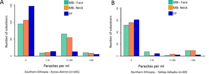
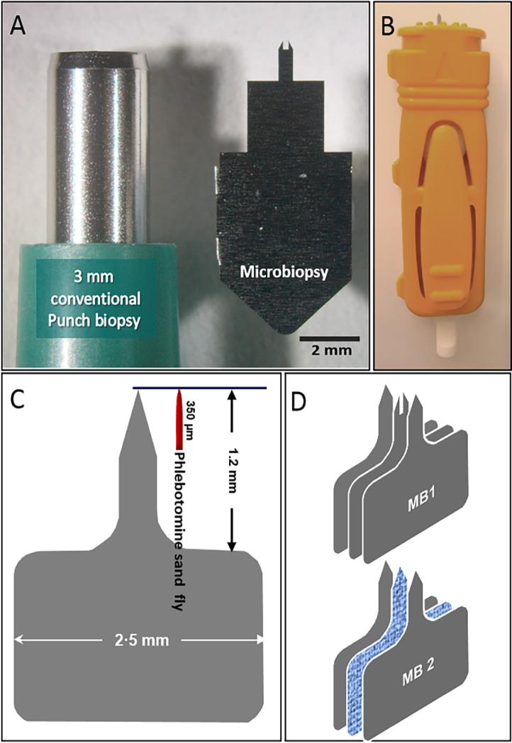
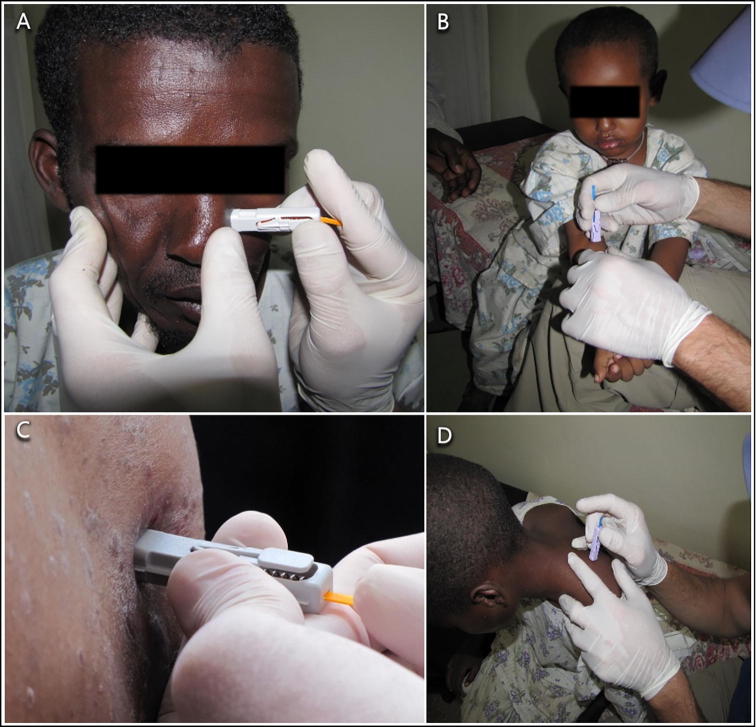
Visceral leishmaniasis (VL) is a potentially lethal, sand fly-borne disease caused by protozoan parasites belonging to the Leishmania donovani species complex. There are several adequate methods for diagnosing VL, but the majority of infected individuals remain asymptomatic, comprising potential parasite reservoirs for transmission of the disease. The gold standard for assessing host infectiousness to biting vector insects is xenodiagnosis (i.e. scoring infection rates among insectary-reared insects that had fed on humans suspected of being infected). However, when it comes to sand flies and leishmaniasis, xenodiagnosis is an intricate operation burdened by logistical hurdles and ethical concerns that prevent its effective application for mass screening of widely dispersed communities, particularly in rural regions of underdeveloped countries. Minimally invasive microbiopsy (MB) devices were designed to penetrate the skin to a depth of ∼200µm and absorb blood as well as skin cell lysates, mimicking the mode by which phlebotomine sand flies acquire blood meals, as well as their composition. MBs taken from 137 of 262 volunteers, living in endemic VL foci in Ethiopia, detected Leishmania parasites that could potentially be imbibed by feeding sand flies. Although the volume of MBs was 10-fold smaller than finger-prick blood samples, Leishmania DNA detection rates from MBs were significantly higher, implying that skin, more often than blood, was the source of parasites. Volunteers with histories of VL were almost as likely as healthy volunteers to test positive by MBs (southern Ethiopian focus: 95% CI: 0.35-2.59, P=1.0. northern Ethiopian focus 0.87: 95% CI: 0.22-3.76, P=1), suggesting the importance of asymptomatic patients as reservoirs of L. donovani. Minimally invasive, painless MBs should be considered for reliably and efficiently evaluating both L. donovani infection rates among large numbers of asymptomatic carriers and their infectiousness to blood-feeding sand flies.

摘要

内脏利什曼病(VL)是一种潜在致命的沙蝇传播疾病,由属于利什曼原虫复合体的原生动物寄生虫引起。有几种方法可以诊断 VL,但大多数受感染的个体仍然无症状,构成了疾病传播的潜在寄生虫储存库。评估宿主对吸血昆虫传播媒介的感染性的金标准是异种诊断(即对在昆虫饲养室中饲养的昆虫进行评分,这些昆虫曾吸食过疑似感染的人类)。然而,对于沙蝇和利什曼病来说,异种诊断是一项复杂的操作,受到后勤障碍和伦理问题的困扰,这些问题阻碍了其在广泛分布的社区中进行大规模筛查的有效应用,特别是在欠发达国家的农村地区。微创微生物活检(MB)装置旨在穿透皮肤深度约 200µm,并吸收血液和皮肤细胞裂解物,模拟白蛉沙蝇获取血餐的方式及其组成。从生活在埃塞俄比亚内脏利什曼病流行地区的 262 名志愿者中的 137 人采集的 MB 检测到了潜在可被喂食沙蝇吸收的利什曼寄生虫。尽管 MB 的体积比指压血样小 10 倍,但从 MB 中检测到利什曼 DNA 的比例要高得多,这意味着皮肤比血液更有可能成为寄生虫的来源。有 VL 病史的志愿者与健康志愿者一样,通过 MB 检测呈阳性的可能性几乎相同(埃塞俄比亚南部焦点:95%CI:0.35-2.59,P=1.0;埃塞俄比亚北部焦点:0.87:95%CI:0.22-3.76,P=1),这表明无症状患者作为 L. donovani 储存库的重要性。微创、无痛的 MB 应该被认为是可靠和有效地评估大量无症状携带者中的 L. donovani 感染率及其对吸血沙蝇的传染性的方法。

https://cdn.ncbi.nlm.nih.gov/pmc/blobs/c1d6/5596977/1d0852a7dee0/gr4.jpg
https://cdn.ncbi.nlm.nih.gov/pmc/blobs/c1d6/5596977/026fdb05ab54/fx1.jpg
https://cdn.ncbi.nlm.nih.gov/pmc/blobs/c1d6/5596977/62b81dc3b22f/gr1.jpg
https://cdn.ncbi.nlm.nih.gov/pmc/blobs/c1d6/5596977/2082e5387697/gr2.jpg
https://cdn.ncbi.nlm.nih.gov/pmc/blobs/c1d6/5596977/d36f8ff8eef6/gr3.jpg
https://cdn.ncbi.nlm.nih.gov/pmc/blobs/c1d6/5596977/1d0852a7dee0/gr4.jpg

相似文献

1
Minimally invasive microbiopsies: a novel sampling method for identifying asymptomatic, potentially infectious carriers of Leishmania donovani.微创微生物活检:一种新型采样方法,用于鉴定无症状、潜在感染利什曼原虫的携带者。
Int J Parasitol. 2017 Sep;47(10-11):609-616. doi: 10.1016/j.ijpara.2017.02.005. Epub 2017 Apr 26.
2
Xenodiagnosis of Leishmania donovani in BALB/c mice using Phlebotomus orientalis: a new laboratory model.利用东方白蛉对BALB/c小鼠进行杜氏利什曼原虫的异种诊断:一种新的实验室模型
Parasit Vectors. 2015 Mar 14;8:158. doi: 10.1186/s13071-015-0765-x.
3
Detection of Leishmania donovani and L. tropica in Ethiopian wild rodents.埃塞俄比亚野生啮齿动物中杜氏利什曼原虫和热带利什曼原虫的检测。
Acta Trop. 2015 May;145:39-44. doi: 10.1016/j.actatropica.2015.02.006. Epub 2015 Feb 18.
4
Evaluation of PCR procedures for detecting and quantifying Leishmania donovani DNA in large numbers of dried human blood samples from a visceral leishmaniasis focus in Northern Ethiopia.评价聚合酶链反应(PCR)程序,以检测和定量来自埃塞俄比亚北部内脏利什曼病流行区大量干燥人血样本中的杜氏利什曼原虫 DNA。
BMC Infect Dis. 2013 Mar 27;13:153. doi: 10.1186/1471-2334-13-153.
5
Quantifying the Infectiousness of Post-Kala-Azar Dermal Leishmaniasis Toward Sand Flies.定量评估卡拉巴肿后皮肤利什曼病对沙蝇的感染性。
Clin Infect Dis. 2019 Jul 2;69(2):251-258. doi: 10.1093/cid/ciy891.
6
Livestock and rodents within an endemic focus of Visceral Leishmaniasis are not reservoir hosts for Leishmania donovani.内脏利什曼病地方性流行区的家畜和啮齿动物不是杜氏利什曼原虫的储存宿主。
PLoS Negl Trop Dis. 2022 Oct 20;16(10):e0010347. doi: 10.1371/journal.pntd.0010347. eCollection 2022 Oct.
7
Role of asymptomatic and symptomatic humans as reservoirs of visceral leishmaniasis in a Mediterranean context.无症状和有症状的人类在 Mediterranean 环境中作为内脏利什曼病储主的作用。
PLoS Negl Trop Dis. 2020 Apr 23;14(4):e0008253. doi: 10.1371/journal.pntd.0008253. eCollection 2020 Apr.
8
Increased Transmissibility of Leishmania donovani From the Mammalian Host to Vector Sand Flies After Multiple Exposures to Sand Fly Bites.多次暴露于白蛉叮咬后,杜氏利什曼原虫从哺乳动物宿主向媒介白蛉的传播能力增强。
J Infect Dis. 2017 Apr 15;215(8):1285-1293. doi: 10.1093/infdis/jix115.
9
First detection of Leishmania donovani in sand flies from Cameroon and its epidemiological implications.首次在喀麦隆的沙蝇中检测到杜氏利什曼原虫及其流行病学意义。
Trop Med Int Health. 2018 Sep;23(9):1014-1021. doi: 10.1111/tmi.13123. Epub 2018 Jul 30.
10
Xenodiagnosis on dogs with visceral leishmaniasis: Canine and sand fly aspects related to the parasite transmission.对内脏利什曼病犬进行异种诊断:与寄生虫传播相关的犬类和白蛉方面
Vet Parasitol. 2016 Jun 15;223:120-6. doi: 10.1016/j.vetpar.2016.04.031. Epub 2016 Apr 22.

引用本文的文献

1
Infectiousness of Leishmania major to Phlebotomus papatasi: differences between natural reservoir host Meriones shawi and laboratory model BALB/c mice.硕大利什曼原虫对巴氏白蛉的感染性:自然宿主肖氏子午沙鼠与实验室模型BALB/c小鼠之间的差异。
PLoS Negl Trop Dis. 2025 Jun 16;19(6):e0013183. doi: 10.1371/journal.pntd.0013183. eCollection 2025 Jun.
2
Safety and reactogenicity of a controlled human infection model of sand fly-transmitted cutaneous leishmaniasis.沙蝇传播的皮肤利什曼病人体感染模型的安全性和反应原性。
Nat Med. 2024 Nov;30(11):3150-3162. doi: 10.1038/s41591-024-03146-9. Epub 2024 Aug 2.
3
Chronic High-Level Parasitemia in HIV-Infected Individuals With or Without Visceral Leishmaniasis in an Endemic Area in Northwest Ethiopia: Potential Superspreaders?

本文引用的文献

1
Transmission Dynamics of Visceral Leishmaniasis in the Indian Subcontinent - A Systematic Literature Review.印度次大陆内脏利什曼病的传播动力学——一项系统文献综述
PLoS Negl Trop Dis. 2016 Aug 4;10(8):e0004896. doi: 10.1371/journal.pntd.0004896. eCollection 2016 Aug.
2
Optimization of loop-mediated isothermal amplification (LAMP) assays for the detection of Leishmania DNA in human blood samples.用于检测人血样本中利什曼原虫DNA的环介导等温扩增(LAMP)检测方法的优化。
Acta Trop. 2016 Oct;162:20-26. doi: 10.1016/j.actatropica.2016.06.009. Epub 2016 Jun 8.
3
New Insights Into the Transmissibility of Leishmania infantum From Dogs to Sand Flies: Experimental Vector-Transmission Reveals Persistent Parasite Depots at Bite Sites.
慢性高水平寄生虫血症在艾滋病毒感染个体中,无论是否存在内脏利什曼病,在埃塞俄比亚西北部的一个流行地区:潜在的超级传播者?
Clin Infect Dis. 2024 Jul 19;79(1):240-246. doi: 10.1093/cid/ciae002.
4
Tape-disc-loop-mediated isothermal amplification (TD-LAMP) method as noninvasive approach for diagnosis of cutaneous leishmaniasis caused by .胶带-圆盘-环介导等温扩增(TD-LAMP)法作为诊断由……引起的皮肤利什曼病的非侵入性方法。 (注:原文中“caused by”后内容缺失)
Heliyon. 2023 Oct 29;9(11):e21397. doi: 10.1016/j.heliyon.2023.e21397. eCollection 2023 Nov.
5
Asymptomatic Leishmania donovani infection and associated factors among blood donors attending at Metema district Blood Bank, Northwest Ethiopia: a cross- sectional study.埃塞俄比亚西北部梅特马地区血库无偿献血者中无症状杜氏利什曼原虫感染及其相关因素:一项横断面研究
Arch Public Health. 2023 Apr 21;81(1):62. doi: 10.1186/s13690-023-01082-7.
6
Visceral Leishmaniasis and the Skin: Dermal Parasite Transmission to Sand Flies.内脏利什曼病与皮肤:皮肤寄生虫向白蛉的传播。
Pathogens. 2022 May 24;11(6):610. doi: 10.3390/pathogens11060610.
7
Spatial Point Pattern Analysis Identifies Mechanisms Shaping the Skin Parasite Landscape in Infection.空间点格局分析确定了在 感染中塑造皮肤寄生虫景观的机制。
Front Immunol. 2021 Dec 16;12:795554. doi: 10.3389/fimmu.2021.795554. eCollection 2021.
8
Diversity and Within-Host Evolution of Leishmania donovani from Visceral Leishmaniasis Patients with and without HIV Coinfection in Northern Ethiopia.来自埃塞俄比亚北部有和无 HIV 共感染内脏利什曼病患者的利什曼原虫多样性和体内进化。
mBio. 2021 Jun 29;12(3):e0097121. doi: 10.1128/mBio.00971-21.
9
Evaluation of qPCR on blood and skin microbiopsies, peripheral blood buffy coat smear, and urine antigen ELISA for diagnosis and test of cure for visceral leishmaniasis in HIV-coinfected patients in India: a prospective cohort study.印度HIV合并感染患者内脏利什曼病诊断及治疗检测中,对血液和皮肤活检组织、外周血 Buffy 层涂片及尿液抗原酶联免疫吸附测定进行定量聚合酶链反应评估:一项前瞻性队列研究。
BMJ Open. 2021 Apr 30;11(4):e042519. doi: 10.1136/bmjopen-2020-042519.
10
Assessing Skin Parasite Load: A Proof of Concept Study of a Microbiopsy Device in an Indian Setting.评估皮肤寄生虫载量:印度环境中一种微创活检设备的概念验证研究。
Front Cell Infect Microbiol. 2021 Mar 11;11:645121. doi: 10.3389/fcimb.2021.645121. eCollection 2021.
婴儿利什曼原虫从狗传播至白蛉的新见解:实验性媒介传播揭示了叮咬部位存在持久性寄生虫库。
J Infect Dis. 2016 Jun 1;213(11):1752-61. doi: 10.1093/infdis/jiw022. Epub 2016 Jan 14.
4
Physiological Age Structure and Leishmania spp. Detection in Phlebotomus (Larroussius) orientalis (Parrot, 1936) (Diptera: Psychodidae) at an Endemic Focus of Visceral Leishmaniasis in Northern Ethiopia.埃塞俄比亚北部内脏利什曼病流行区东方白蛉(1936年,帕罗特)(双翅目:毛蠓科)的生理年龄结构与利什曼原虫属检测
J Trop Med. 2015;2015:710528. doi: 10.1155/2015/710528. Epub 2015 Jul 29.
5
Eco-epidemiology of visceral leishmaniasis in Ethiopia.埃塞俄比亚内脏利什曼病的生态流行病学
Parasit Vectors. 2015 Jul 19;8:381. doi: 10.1186/s13071-015-0987-y.
6
Comparison of Bloodmeal Digestion and the Peritrophic Matrix in Four Sand Fly Species Differing in Susceptibility to Leishmania donovani.四种对杜氏利什曼原虫易感性不同的白蛉物种血餐消化与围食膜的比较
PLoS One. 2015 Jun 1;10(6):e0128203. doi: 10.1371/journal.pone.0128203. eCollection 2015.
7
Quantifying the contribution of hosts with different parasite concentrations to the transmission of visceral leishmaniasis in Ethiopia.量化埃塞俄比亚不同寄生虫浓度宿主对内脏利什曼病传播的贡献。
PLoS Negl Trop Dis. 2014 Oct 30;8(10):e3288. doi: 10.1371/journal.pntd.0003288. eCollection 2014 Oct.
8
Microbiopsy engineered for minimally invasive and suture-free sub-millimetre skin sampling.为实现微创和免缝合的亚毫米级皮肤采样而设计的微型活检技术。
F1000Res. 2013 May 2;2:120. doi: 10.12688/f1000research.2-120.v2. eCollection 2013.
9
Cutaneous leishmaniasis in North Africa: a review.北非的皮肤利什曼病:综述
Parasite. 2014;21:14. doi: 10.1051/parasite/2014014. Epub 2014 Mar 14.
10
Asymptomatic Leishmania infection: a new challenge for Leishmania control.无症状利什曼原虫感染:利什曼原虫控制面临的新挑战。
Clin Infect Dis. 2014 May;58(10):1424-9. doi: 10.1093/cid/ciu102. Epub 2014 Feb 27.