Yuan Jianmin, Makris Gregory, Patterson Andrew, Usman Ammara, Das Tilak, Priest Andrew, Teng Zhongzhao, Hilborne Sarah, Prudencio Dario, Gillard Jonathan, Graves Martin
Department of Radiology, University of Cambridge, Cambridge, UK.
Department of Radiology, Cambridge University Hospitals NHS Foundation Trust, Cambridge, UK.
MAGMA. 2018 Feb;31(1):191-199. doi: 10.1007/s10334-017-0621-4. Epub 2017 Apr 28.
This study aims to explore the relationship between plaque surface morphology and neovascularization using a high temporal and spatial resolution 4D contrast-enhanced MRI/MRA sequence.
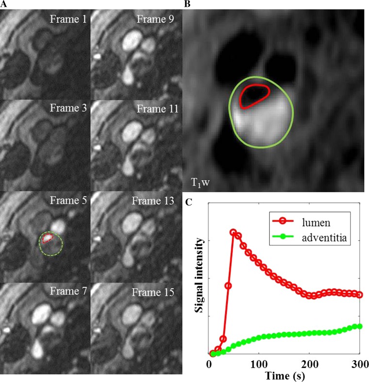
Twenty one patients with either recent symptoms or a carotid artery stenosis ≥40% were recruited in this study. Plaque surface morphology and luminal stenosis were determined from the arterial phase MRA images. Carotid neovascularization was evaluated by a previously validated pharmacokinetic (PK) modeling approach. K (transfer constant) and v (partial plasma volume) were calculated in both the adventitia and plaque.
Image acquisition and analysis was successfully performed in 28 arteries. Mean luminal stenosis was 44% (range 11-82%). Both adventitial and plaque K in ulcerated/irregular plaques were significantly higher than smooth plaques (0.079 ± 0.018 vs. 0.064 ± 0.011 min, p = 0.02; 0.065 ± 0.013 vs. 0.055 ± 0.010 min, p = 0.03, respectively). Positive correlations between adventitial K and v against stenosis were observed (r = 0.44, p = 0.02; r = 0.55, p = 0.01, respectively).
This study demonstrates the feasibility of using a single sequence to acquire both high resolution 4D CE-MRA and DCE-MRI to evaluate both plaque surface morphology and function. The results demonstrate significant relationships between lumen surface morphology and neovascularization.
本研究旨在使用高时间和空间分辨率的4D对比增强MRI/MRA序列,探讨斑块表面形态与新生血管形成之间的关系。
本研究招募了21例近期有症状或颈动脉狭窄≥40%的患者。从动脉期MRA图像确定斑块表面形态和管腔狭窄情况。采用先前验证的药代动力学(PK)建模方法评估颈动脉新生血管形成。在外膜和斑块中计算K(转运常数)和v(部分血浆容积)。
成功对28条动脉进行了图像采集和分析。平均管腔狭窄率为44%(范围11 - 82%)。溃疡/不规则斑块的外膜和斑块K均显著高于光滑斑块(分别为0.079±0.018对0.064±0.011分钟,p = 0.02;0.065±0.013对0.055±0.010分钟,p = 0.03)。观察到外膜K和v与狭窄之间呈正相关(分别为r = 0.44,p = 0.02;r = 0.55,p = 0.01)。
本研究证明了使用单一序列同时采集高分辨率4D CE-MRA和DCE-MRI以评估斑块表面形态和功能的可行性。结果表明管腔表面形态与新生血管形成之间存在显著关系。