Michelotto Ennio, Tarantino Nicola, Ostuni Vittoria, Pedote Pasquale, Colonna Paolo, Guglielmi Riccardo
Department of Cardiology, Hospital Policlinico of Bari, Bari, Italy.
Department of Radiology, Hospital Policlinico of Bari, Bari, Italy.
J Cardiovasc Echogr. 2013 Oct-Dec;23(4):106-110. doi: 10.4103/2211-4122.127412.
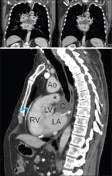
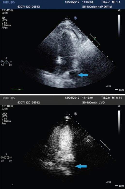
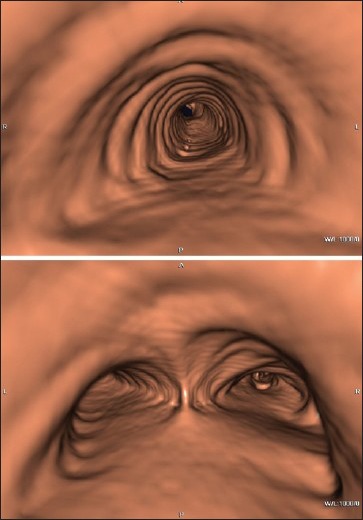
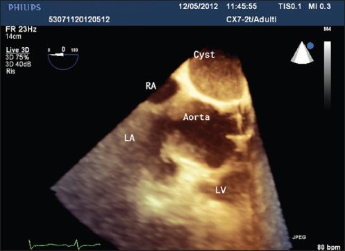
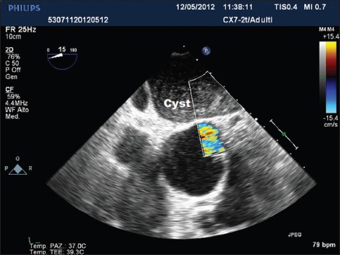
We report a case of a 76-year-old man, with the occasional finding of a mediastinal cyst because of subtle chronic dysphagia associated to sore throat, belching, and dysphonia. The paraesophageal cyst in the central mediastinum was studied with computed tomography (CT) scan and transesophageal three-dimensional (3D) echocardiography with contrast echo. In order to clarify doubts about localization (intra- versus extrapericardial) of the mediastinal cystic lesion the 3D transesophageal echocardiography (TEE) confirmed the presence of a large round cystic mass located contiguous to the esophagus, the left atrium and the aortic root/pulmonary trunk (located at the front of the lesion), as well as located intrapericardial. The cystic mass showed no blood flow at color Doppler mode and at ultrasound contrast echo with SonoVue agent. Due to the paucity of symptoms and to the definite imaging information of this intrapericardial cyst of nonvascular nature, due to pericardial cyst in an extremely unusual location, surgery was not performed. At follow-up of 1 month echocardiogram and 3 month CT scan the cyst appeared unchanged in dimensions.
我们报告一例76岁男性患者,因与咽痛、嗳气和声音嘶哑相关的轻微慢性吞咽困难偶然发现纵隔囊肿。对位于纵隔中部的食管旁囊肿进行了计算机断层扫描(CT)和经食管三维(3D)超声心动图造影检查。为了明确纵隔囊性病变的定位(心包内还是心包外)疑问,三维经食管超声心动图(TEE)证实存在一个大的圆形囊性肿块,与食管、左心房和主动脉根部/肺动脉主干相邻(位于病变前方),且位于心包内。囊性肿块在彩色多普勒模式下及使用声诺维造影剂的超声造影检查中均未显示血流信号。由于症状轻微且该非血管性心包内囊肿的影像学信息明确,且囊肿位置极为罕见,故未进行手术。在1个月的超声心动图随访和3个月的CT扫描中,囊肿大小未见变化。