Kato Tatsuya, Toyooka Tatsushi, Ibuki Yuko, Masuda Shuichi, Watanabe Masatoshi, Totsuka Yukari
Division of Carcinogenesis and Cancer Prevention, National Cancer Center Research Institute, 1-1 Tsukiji 5-chome, Chuo-ku, Tokyo, 104-0045 Japan.
Graduate School of Food and Nutritional Sciences, University of Shizuoka, 52-1, Yada, Shizuoka 422-8526 Japan.
Genes Environ. 2017 May 1;39:12. doi: 10.1186/s41021-017-0075-y. eCollection 2017.
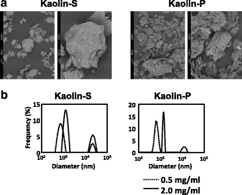
Kaolin is white clay mineral with the chemical composition AlSiO(OH), and many varieties of kaolins having different crystal structures are utilized in industrial, cosmetic and medical fields. To evaluate the effect of physicochemical character differences on the genotoxicity of kaolin, two types of kaolin, kaolin-S with smooth, sphere-shaped crystals, and kaolin-P with clusters of thin pseudohexagonal plates, were used in the study.
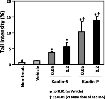
ICR mice were intratracheally instilled with the kaolins (0.05 and 0.2 mg/mouse), and comet assay was performed on their lungs. Both kaolins showed DNA damage in the lungs of the mice, however the DNA damaging potency was much higher with kaolin-P than that with kaolin-S. In order to clarify the mechanisms for the different genotoxic potency, we examined the incorporation rate and ROS generation of these two types of kaolin in alveolar epithelial A549 and macrophage-like RAW264 cells, using flow cytometric (FCM) analysis. Kaolin-P showed a higher incorporation rate into the mammalian cells and ROS generation than that of kaolin-S. Especially, RAW264 cells aggressively incorporated kaolins, and generated ROS, whereas almost no ROS generation was observed in A549 cells. In addition, inflammatory cytokines were quantified, using the ELISA method, to understand further genotoxic potency differences of kaolins. Concentrations of interleukin-1β (IL-1β) and tumor necrosis factor-α (TNF-α) in the media were increased by exposure to both kaolins, but in the case of kaolin-P, these inflammatory cytokines were significantly elevated. Based on these findings, differences of genotoxic potency may contribute to incorporation rates into immune cells. Furthermore, it is likely that immune cells and epithelial cells might closely interact with each other for the appearance of genotoxocity in vivo. In order to clarify the interaction between epithelial and immune cells, A549 and RAW264 were co-cultured and RAW264 cells only were exposed to kaolins, then subsequently A549 was applied to FCM analysis and comet assay. DNA damage observed in the A549 cells markedly increased in the presence of kaolin-exposed RAW264 cells compared to the single culture.
From these observations, it is suggested that mechanisms of kaolin genotoxicity against epithelial cells are through the activation of macrophage cells. Therefore, it is thought that interactions between epithelial and immune cells would be very important for evaluation of the genotoxicity of fine particulate matter. We also showed here that co-culture models of epithelial and immune cells could be used as suitable models for evaluation of lung genotoxicity of fine particulate matter, including nanomaterials, as in vivo mimicking systems.
高岭土是一种化学成分 为AlSiO(OH)的白色粘土矿物,多种具有不同晶体结构的高岭土被应用于工业、化妆品和医学领域。为了评估物理化学性质差异对高岭土遗传毒性的影响,本研究使用了两种类型的高岭土:具有光滑球形晶体的高岭土-S和具有薄假六边形板簇的高岭土-P。
将高岭土(0.05和0.2毫克/只小鼠)经气管内滴注到ICR小鼠体内,并对其肺部进行彗星试验。两种高岭土均在小鼠肺部显示出DNA损伤,然而高岭土-P的DNA损伤能力比高岭土-S高得多。为了阐明不同遗传毒性效力的机制,我们使用流式细胞术(FCM)分析检测了这两种高岭土在肺泡上皮A549细胞和巨噬细胞样RAW264细胞中的摄取率和活性氧生成情况。高岭土-P在哺乳动物细胞中的摄取率和活性氧生成均高于高岭土-S。特别是,RAW264细胞能大量摄取高岭土并产生活性氧,而在A549细胞中几乎未观察到活性氧生成。此外,使用酶联免疫吸附测定(ELISA)方法对炎性细胞因子进行定量,以进一步了解高岭土遗传毒性效力的差异。两种高岭土暴露均使培养基中白细胞介素-1β(IL-1β)和肿瘤坏死因子-α(TNF-α)的浓度升高,但在高岭土-P的情况下,这些炎性细胞因子显著升高。基于这些发现,遗传毒性效力的差异可能与免疫细胞的摄取率有关。此外,免疫细胞和上皮细胞在体内可能密切相互作用导致遗传毒性的出现。为了阐明上皮细胞和免疫细胞之间的相互作用,将A549细胞和RAW264细胞共培养,仅将RAW264细胞暴露于高岭土,随后对A549细胞进行FCM分析和彗星试验。与单一培养相比,在存在暴露于高岭土的RAW264细胞的情况下,A549细胞中观察到的DNA损伤明显增加。
从这些观察结果来看,提示高岭土对上皮细胞的遗传毒性机制是通过巨噬细胞的激活。因此,认为上皮细胞和免疫细胞之间的相互作用对于评估细颗粒物的遗传毒性非常重要。我们在此还表明,上皮细胞和免疫细胞的共培养模型可作为评估包括纳米材料在内的细颗粒物肺遗传毒性的合适模型,作为体内模拟系统。