Wang Shaohui, Wang Ximing, Boone Jasmine, Wie Jin, Yip Kay-Pong, Zhang Jie, Wang Lei, Liu Ruisheng
Department of Molecular Pharmacology & Physiology, University of South Florida Morsani College of Medicine, Tampa, Florida, USA.
Present Address: Shandong Medical Imaging Research Institute, Shandong provincial key laboratory of diagnosis and treatment of cardio-cerebral vascular disease, Shandong University, Jinan, China.
Kidney Blood Press Res. 2017;42(2):220-231. doi: 10.1159/000476018. Epub 2017 May 4.
BACKGROUND/AIMS: The hanging drop technique is a well-established method used in culture of animal tissues. However, this method has not been used in adult kidney tissue culture yet. This study was to explore the feasibility of using this technique for culturing adult kidney cortex to study the time course of RNA viability in the tubules and vasculature, as well as the tissue structural integrity.
In each Petri dish with the plate covered with sterile buffer, a section of mouse renal cortex was cultured within a drop of DMEM culture medium on the inner surface of the lip facing downward. The tissue were then harvested at each specific time points for Real-time PCR analysis and histological studies.
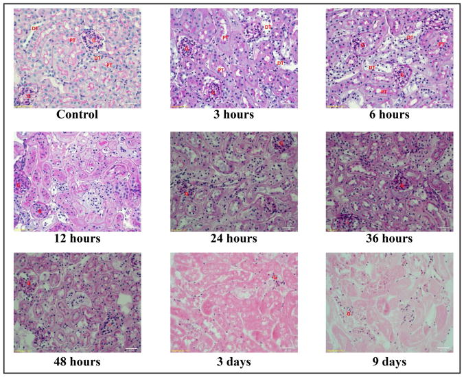
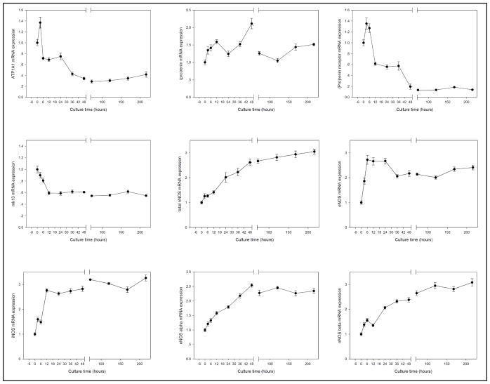
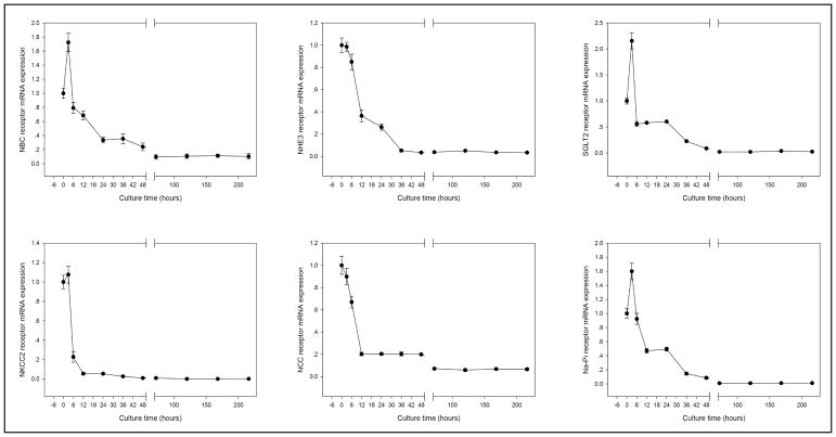
The results showed that the mRNA level of most Na+ related transporters and cotransporters were stably maintained within 6 hours in culture, and that the mRNA level of most receptors found in the vasculature and glomeruli were stably maintained for up to 9 days in culture. Paraffin sections of the cultured renal cortex indicated that the tubules began to lose tubular integrity after 6 hours, but the glomeruli and vasculatures were still recognizable up to 9 days in culture.
We concluded that adult kidney tissue culture by hanging drop method can be used to study gene expressions in vasculature and glomeruli.
背景/目的:悬滴技术是动物组织培养中一种成熟的方法。然而,该方法尚未用于成年肾组织培养。本研究旨在探讨使用该技术培养成年肾皮质以研究肾小管和血管中RNA活力的时间进程以及组织结构完整性的可行性。
在每个覆盖有无菌缓冲液的培养皿中,将一段小鼠肾皮质培养在向下的唇部内表面的一滴DMEM培养基中。然后在每个特定时间点收获组织用于实时PCR分析和组织学研究。
结果表明,大多数与Na+相关的转运体和协同转运体的mRNA水平在培养6小时内稳定维持,并且在血管和肾小球中发现的大多数受体的mRNA水平在培养长达9天内稳定维持。培养的肾皮质石蜡切片表明,肾小管在6小时后开始失去肾小管完整性,但肾小球和血管在培养9天内仍可识别。
我们得出结论,通过悬滴法进行成年肾组织培养可用于研究血管和肾小球中的基因表达。