Chen X W, Yu T J, Zhang J, Li Y, Chen H L, Yang G F, Yu W, Liu Y Z, Liu X X, Duan C F, Tang H L, Qiu M, Wang C L, Zheng H, Yue J, Guo A M, Yang J
Department of Pharmacology, School of Basic Medical Sciences, Wuhan University, Wuhan, China.
Animal Experimental Center of Wuhan University, Wuhan, China.
Oncogene. 2017 Aug 31;36(35):5045-5057. doi: 10.1038/onc.2017.118. Epub 2017 May 8.
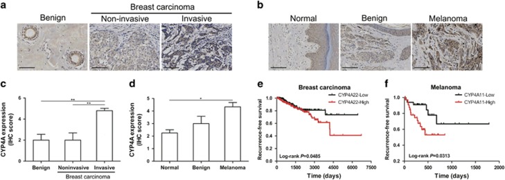
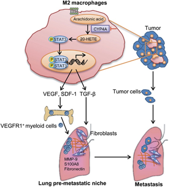
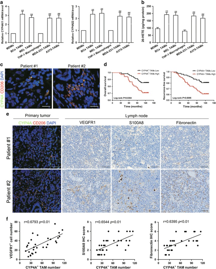
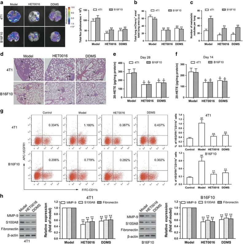
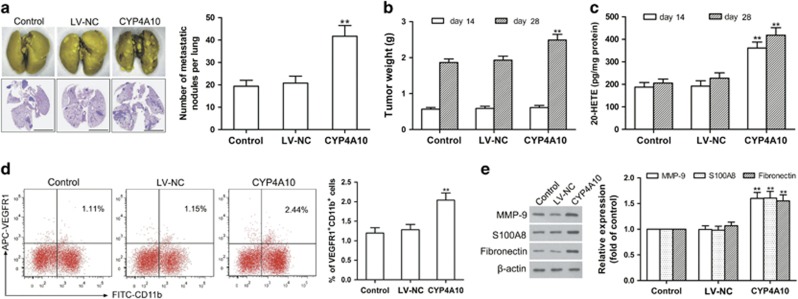
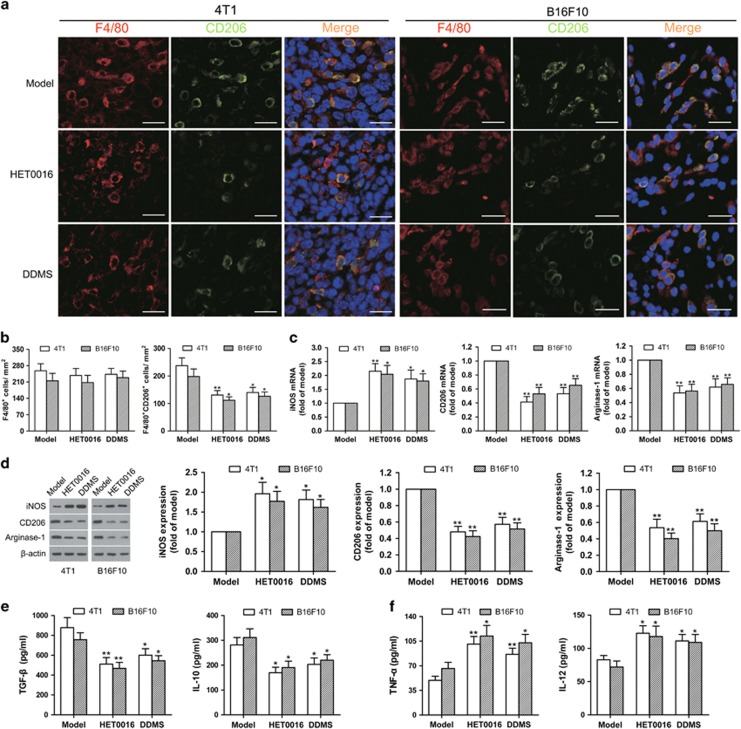
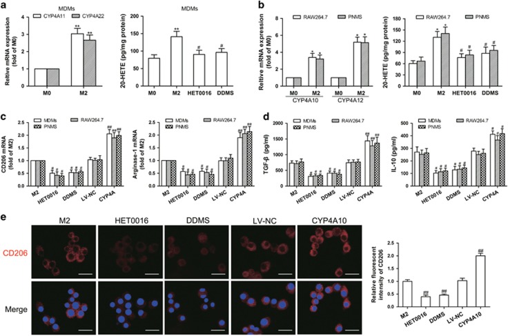
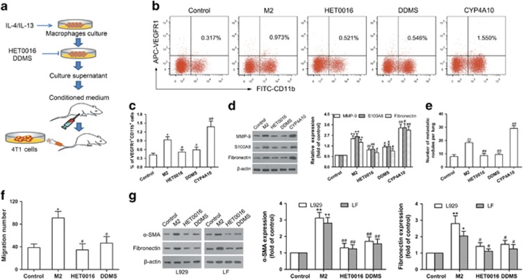
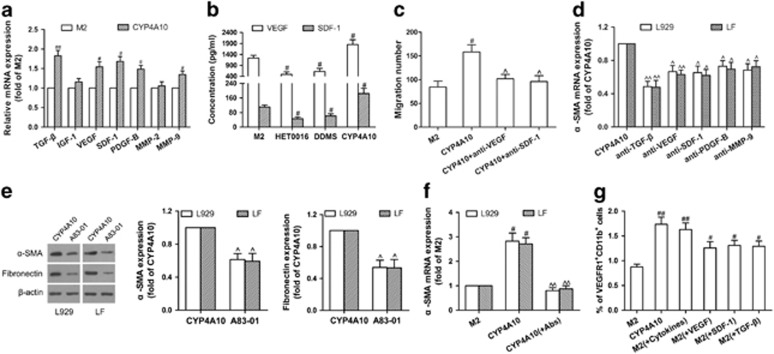
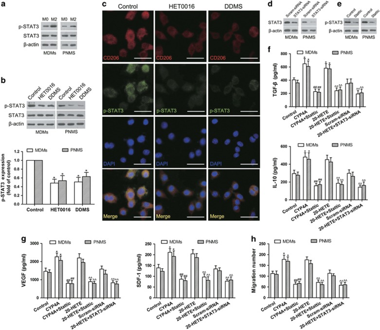
Tumor-associated macrophages (TAMs) play an essential role in metastasis. However, what enables TAMs to have a superior capacity to establish pre-metastatic microenvironment in distant organs is unclear. Here we have begun to uncover the effects of cytochrome P450 (CYP) 4A in TAMs on lung pre-metastatic niche formation and metastasis. CYP4A TAM infiltration was positively associated with metastasis, pre-metastatic niche formation and poor prognosis in breast cancer patients. The pharmacological inhibition of CYP4A reduced lung pre-metastatic niche formation (evidenced by a decrease in vascular endothelial growth factor receptor 1 positive (VEGFR1) myeloid cell recruitment and pro-metastatic protein expression) and metastatic burden, accompanied with TAM polarization away from the M2 phenotype in spontaneous metastasis models of 4T1 breast cancer and B16F10 melanoma. Co-implantation of 4T1 cells with CYP4A10 macrophages promoted lung pre-metastatic niche formation and metastasis. Depletion of TAMs disrupted lung pre-metastatic niches and thereby prevented metastasis. Treatment with the CM from CYP4A10 M2 macrophages (M2) increased pre-metastatic niche formation and metastatic burden in the lungs, whereas CYP4A inhibition attenuated these effects. In vitro TAM polarization away from the M2 phenotype induced by CYP4A inhibition decreased VEGFR1 myeloid cell migration and fibronectin expression, accompanied with downregulation of STAT3 signaling. Conversely, overexpression of CYP4A or exogenous addition of 20-hydroxyeicosatetraenoic acid promoted M2 polarization and cytokine production of macrophages and thereby enhanced migration of VEGFR1 myeloid cells, which were reversed by siRNA or pharmacological inhibition of STAT3. Importantly, a combined blocking M2 macrophage-derived factors TGF-β, VEGF and SDF-1 abolished VEGFR1 myeloid cell migration and fibroblast activation induced by CYP4A. In summary, CYP4A in TAMs is crucial for lung pre-metastatic niche formation and metastasis, and may serve as a potential therapeutic target in human cancer.
肿瘤相关巨噬细胞(TAM)在转移过程中发挥着重要作用。然而,尚不清楚是什么使TAM具有在远处器官建立转移前微环境的卓越能力。在此,我们开始揭示细胞色素P450(CYP)4A在TAM中对肺转移前生态位形成和转移的影响。CYP4A在TAM中的浸润与乳腺癌患者的转移、转移前生态位形成及不良预后呈正相关。CYP4A的药理抑制作用减少了肺转移前生态位的形成(表现为血管内皮生长因子受体1阳性(VEGFR1)髓样细胞募集和促转移蛋白表达的减少)以及转移负担,同时在4T1乳腺癌和B16F10黑色素瘤的自发转移模型中,TAM向远离M2表型的方向极化。将4T1细胞与CYP4A10巨噬细胞共同植入促进了肺转移前生态位的形成和转移。TAM的清除破坏了肺转移前生态位,从而阻止了转移。用来自CYP4A10 M2巨噬细胞(M2)的条件培养基(CM)处理可增加肺转移前生态位的形成和转移负担,而CYP4A抑制可减弱这些作用。体外实验中,CYP4A抑制诱导的TAM向远离M2表型的极化减少了VEGFR1髓样细胞迁移和纤连蛋白表达,同时伴随着信号转导和转录激活因子3(STAT3)信号的下调。相反,CYP4A的过表达或外源性添加20-羟基二十碳四烯酸促进了巨噬细胞的M2极化和细胞因子产生,从而增强了VEGFR1髓样细胞的迁移,而小干扰RNA(siRNA)或STAT3的药理抑制可逆转这种作用。重要的是,联合阻断M2巨噬细胞衍生因子转化生长因子-β(TGF-β)、血管内皮生长因子(VEGF)和基质细胞衍生因子-1(SDF-1)可消除CYP4A诱导的VEGFR1髓样细胞迁移和成纤维细胞活化。总之,TAM中的CYP4A对肺转移前生态位的形成和转移至关重要,可能成为人类癌症的潜在治疗靶点。