Baselet Bjorn, Belmans Niels, Coninx Emma, Lowe Donna, Janssen Ann, Michaux Arlette, Tabury Kevin, Raj Kenneth, Quintens Roel, Benotmane Mohammed A, Baatout Sarah, Sonveaux Pierre, Aerts An
Radiobiology Unit, Belgian Nuclear Research Centre (SCK•CEN), Institute for Environment, Health and SafetyMol, Belgium.
Institut de Recherche Expérimentale et Clinique (IREC), Pole of Pharmacology & Therapeutics, Université catholique de LouvainBrussels, Belgium.
Front Pharmacol. 2017 Apr 25;8:213. doi: 10.3389/fphar.2017.00213. eCollection 2017.
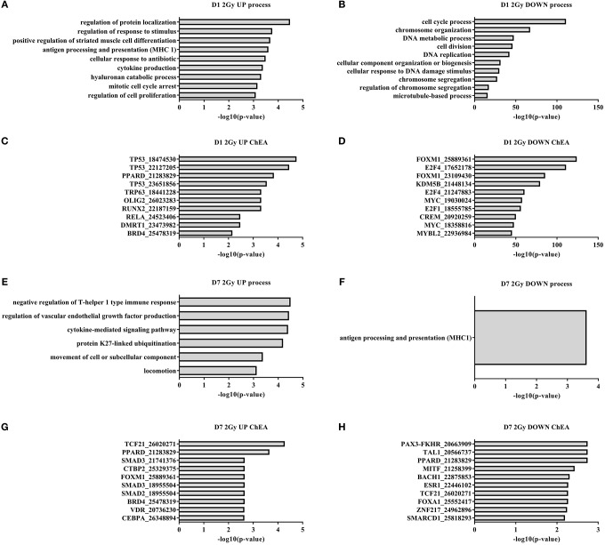
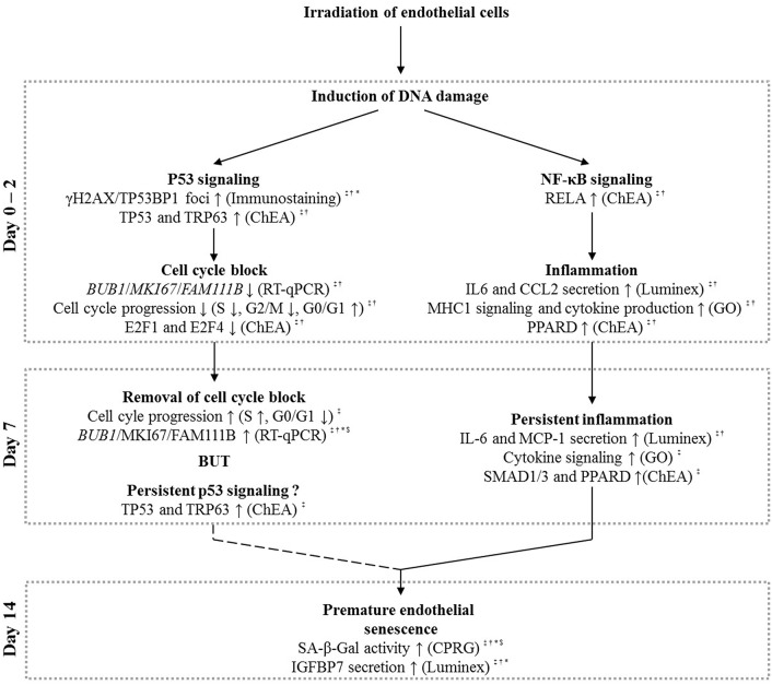
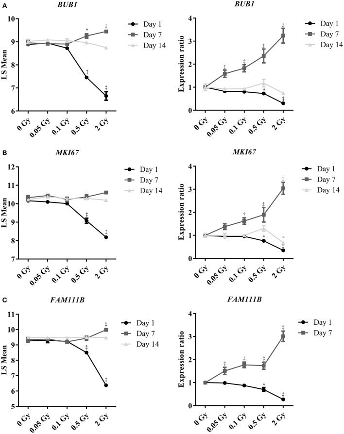
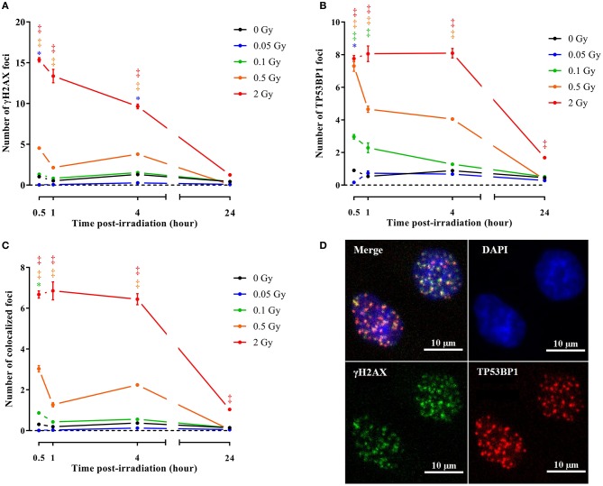
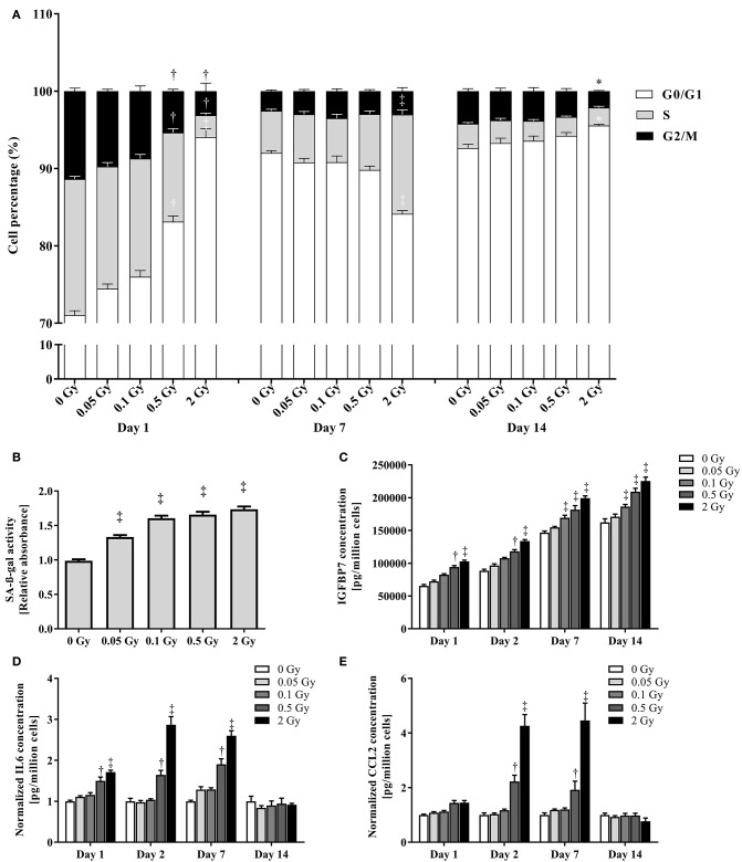
Epidemiological data suggests an excess risk of cardiovascular disease (CVD) at low doses (0.05 and 0.1 Gy) of ionizing radiation (IR). Furthermore, the underlying biological and molecular mechanisms of radiation-induced CVD are still unclear. Because damage to the endothelium could be critical in IR-related CVD, this study aimed to identify the effects of radiation on immortalized endothelial cells in the context of atherosclerosis. Microarrays and RT-qPCR were used to compare the response of endothelial cells irradiated with a single X-ray dose (0.05, 0.1, 0.5, 2 Gy) measured after various post-irradiation (repair) times (1 day, 7 days, 14 days). To consolidate and mechanistically support the endothelial cell response to X-ray exposure identified via microarray analysis, DNA repair signaling (γH2AX/TP53BP1-foci quantification), cell cycle progression (BrdU/7AAD flow cytometric analysis), cellular senescence (β-galactosidase assay with CPRG and IGFBP7 quantification) and pro-inflammatory status (IL6 and CCL2) was assessed. Microarray results indicated persistent changes in cell cycle progression and inflammation. Cells underwent G1 arrest in a dose-dependent manner after high doses (0.5 and 2 Gy), which was compensated by increased proliferation after 1 week and almost normalized after 2 weeks. However, at this point irradiated cells showed an increased β-Gal activity and IGFBP7 secretion, indicative of premature senescence. The production of pro-inflammatory cytokines IL6 and CCL2 was increased at early time points. IR induces pro-atherosclerotic processes in endothelial cells in a dose-dependent manner. These findings give an incentive for further research on the shape of the dose-response curve, as we show that even low doses of IR can induce premature endothelial senescence at later time points. Furthermore, our findings on the time- and dose-dependent response regarding differentially expressed genes, cell cycle progression, inflammation and senescence bring novel insights into the underlying molecular mechanisms of the endothelial response to X-ray radiation. This may in turn lead to the development of risk-reducing strategies to prevent IR-induced CVD, such as the use of cell cycle modulators and anti-inflammatory drugs as radioprotectors and/or radiation mitigators.
流行病学数据表明,低剂量(0.05和0.1 Gy)电离辐射(IR)会增加心血管疾病(CVD)的风险。此外,辐射诱发CVD的潜在生物学和分子机制仍不清楚。由于内皮损伤可能是IR相关CVD的关键因素,本研究旨在确定在动脉粥样硬化背景下辐射对永生化内皮细胞的影响。使用微阵列和RT-qPCR比较单次X射线剂量(0.05、0.1、0.5、2 Gy)照射后不同辐射后(修复)时间(1天、7天、14天)测量的内皮细胞反应。为了巩固并从机制上支持通过微阵列分析确定的内皮细胞对X射线照射的反应,评估了DNA修复信号(γH2AX/TP53BP1灶定量)、细胞周期进程(BrdU/7AAD流式细胞术分析)、细胞衰老(用CPRG进行β-半乳糖苷酶测定和IGFBP7定量)和促炎状态(IL6和CCL2)。微阵列结果表明细胞周期进程和炎症存在持续变化。高剂量(0.5和2 Gy)后细胞以剂量依赖方式发生G1期阻滞,1周后增殖增加可对此进行补偿,2周后几乎恢复正常。然而,此时照射后的细胞显示β-半乳糖苷酶活性增加和IGFBP7分泌增加,表明过早衰老。促炎细胞因子IL6和CCL2的产生在早期时间点增加。IR以剂量依赖方式在内皮细胞中诱导促动脉粥样硬化过程。这些发现为进一步研究剂量反应曲线的形状提供了动力,因为我们表明即使是低剂量的IR在后期也可诱导内皮细胞过早衰老。此外,我们关于差异表达基因、细胞周期进程、炎症和衰老的时间和剂量依赖性反应的发现为内皮细胞对X射线辐射反应的潜在分子机制带来了新的见解。这反过来可能会导致开发降低风险的策略来预防IR诱发的CVD,例如使用细胞周期调节剂和抗炎药物作为辐射防护剂和/或辐射减轻剂。