• 文献检索
  • 文档翻译
  • 深度研究
  • 学术资讯
  • Suppr Zotero 插件Zotero 插件
  • 邀请有礼
  • 套餐&价格
  • 历史记录
应用&插件
Suppr Zotero 插件Zotero 插件浏览器插件Mac 客户端Windows 客户端微信小程序
定价
高级版会员购买积分包购买API积分包
服务
文献检索文档翻译深度研究API 文档MCP 服务
关于我们
关于 Suppr公司介绍联系我们用户协议隐私条款
关注我们

Suppr 超能文献

核心技术专利:CN118964589B侵权必究
粤ICP备2023148730 号-1Suppr @ 2026

文献检索

告别复杂PubMed语法,用中文像聊天一样搜索,搜遍4000万医学文献。AI智能推荐,让科研检索更轻松。

立即免费搜索

文件翻译

保留排版,准确专业,支持PDF/Word/PPT等文件格式,支持 12+语言互译。

免费翻译文档

深度研究

AI帮你快速写综述,25分钟生成高质量综述,智能提取关键信息,辅助科研写作。

立即免费体验

光学相干断层扫描作为威尔逊病患者神经退行性变的标志物

Optical coherence tomography as a marker of neurodegeneration in patients with Wilson's disease.

作者信息

Langwińska-Wośko Ewa, Litwin Tomasz, Dzieżyc Karolina, Karlinski Michał, Członkowska Anna

机构信息

Department of Ophthalmology, Medical University of Warsaw, Warsaw, Poland.

2nd Department of Neurology, Institute of Psychiatry and Neurology, Sobieskiego 9, 02-957, Warsaw, Poland.

出版信息

Acta Neurol Belg. 2017 Dec;117(4):867-871. doi: 10.1007/s13760-017-0788-5. Epub 2017 May 9.

DOI:10.1007/s13760-017-0788-5
PMID:28488258
原文链接:https://pmc.ncbi.nlm.nih.gov/articles/PMC5670193/
Abstract

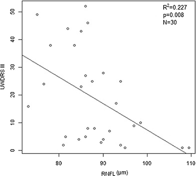
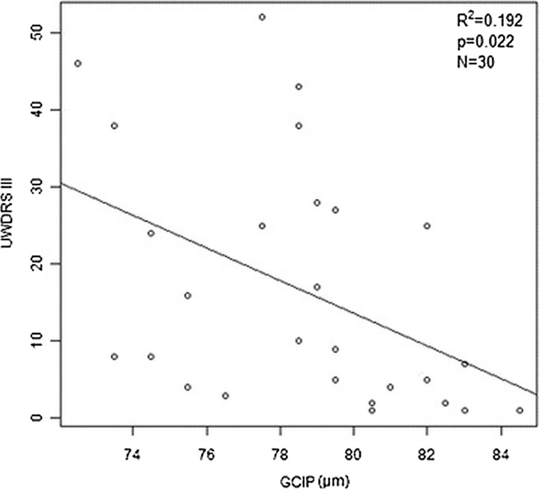
Wilson's disease (WD) is an inherited autosomal recessive disorder that leads to pathological copper accumulation in different organs. Optical coherence tomography (OCT) is proposed as a marker of neurodegeneration in many neurological diseases. Thinning of the total retinal nerve fiber layer (RNFL) and macular thickness (Mth) examined by OCT was detected in patients with WD, especially those with brain magnetic resonance imaging changes. The aim of this study was to evaluate the relationship between OCT parameters and the progression of neurological signs measured by the Unified Wilson's Disease Rating Scale (UWDRS) in patients with WD. Consecutive patients with WD admitted to the Department of Neurology underwent OCT to assess the thickness of the macula and total RNFL. Patients also had neurologic assessments according to the UWDRS part III. Patients were divided into two groups based on the presence (UWDRS+) and absence (UWDRS-) of neurological symptoms. Fifty-eight patients (34 females, 24 males) were enrolled. Mean duration of treatment was 9 years (standard deviation [SD], ±10.8). The mean UWDRS score at the time of study was 8.4 (range 1-52; SD ±13.9) points. Total RNFL as well as macula thickness were significantly decreased in the UWDRS+ group versus the UWDRS- group. A significant negative correlation was found between OCT parameters (RNFL and Mth measurements) and neurological impairment according the UWDRS scale. This study confirms that OCT may be a useful tool for measuring the degree of neurodegeneration in patients with WD, and may play role in monitoring disease progression.

摘要

威尔逊病(WD)是一种常染色体隐性遗传疾病,可导致不同器官出现病理性铜蓄积。光学相干断层扫描(OCT)被提议作为多种神经疾病中神经退行性变的一个标志物。通过OCT检测发现,WD患者存在视网膜神经纤维层(RNFL)总厚度和黄斑厚度(Mth)变薄的情况,尤其是那些有脑磁共振成像改变的患者。本研究的目的是评估WD患者中OCT参数与通过统一威尔逊病评定量表(UWDRS)测量的神经体征进展之间的关系。连续入住神经内科的WD患者接受OCT检查以评估黄斑和RNFL总厚度。患者还根据UWDRS第三部分进行了神经学评估。根据是否存在神经症状,将患者分为两组(UWDRS +组和UWDRS -组)。共纳入58例患者(34例女性,24例男性)。平均治疗时间为9年(标准差[SD],±10.8)。研究时的平均UWDRS评分为8.4分(范围1 - 52;SD±13.9)。与UWDRS -组相比,UWDRS +组的RNFL总厚度以及黄斑厚度均显著降低。根据UWDRS量表,发现OCT参数(RNFL和Mth测量值)与神经功能损害之间存在显著负相关。本研究证实,OCT可能是测量WD患者神经退行性变程度的有用工具,并可能在监测疾病进展中发挥作用。

https://cdn.ncbi.nlm.nih.gov/pmc/blobs/35ca/5670193/c8e69a1a7233/13760_2017_788_Fig2_HTML.jpg
https://cdn.ncbi.nlm.nih.gov/pmc/blobs/35ca/5670193/ebe5542c548d/13760_2017_788_Fig1_HTML.jpg
https://cdn.ncbi.nlm.nih.gov/pmc/blobs/35ca/5670193/c8e69a1a7233/13760_2017_788_Fig2_HTML.jpg
https://cdn.ncbi.nlm.nih.gov/pmc/blobs/35ca/5670193/ebe5542c548d/13760_2017_788_Fig1_HTML.jpg
https://cdn.ncbi.nlm.nih.gov/pmc/blobs/35ca/5670193/c8e69a1a7233/13760_2017_788_Fig2_HTML.jpg

相似文献

1
Optical coherence tomography as a marker of neurodegeneration in patients with Wilson's disease.光学相干断层扫描作为威尔逊病患者神经退行性变的标志物
Acta Neurol Belg. 2017 Dec;117(4):867-871. doi: 10.1007/s13760-017-0788-5. Epub 2017 May 9.
2
Comparative assessment of clinical rating scales in Wilson's disease.威尔逊病临床评定量表的比较评估
BMC Neurol. 2017 Jul 21;17(1):140. doi: 10.1186/s12883-017-0921-3.
3
Optical coherence tomography in patients with Wilson's disease.Wilson 病患者的光学相干断层扫描。
Acta Neurol Scand. 2021 Aug;144(2):149-154. doi: 10.1111/ane.13431. Epub 2021 Apr 21.
4
Retinal neurodegeneration in Wilson's disease revealed by spectral domain optical coherence tomography.Wilson 病的光谱域光学相干断层扫描显示的视网膜神经退行性变。
PLoS One. 2012;7(11):e49825. doi: 10.1371/journal.pone.0049825. Epub 2012 Nov 16.
5
Optical coherence tomography in patients with Wilson's disease.Wilson 病患者的光学相干断层扫描。
Brain Behav. 2023 Jun;13(6):e3014. doi: 10.1002/brb3.3014. Epub 2023 Apr 16.
6
Optical coherence tomography and electrophysiology of retinal and visual pathways in Wilson's disease.威尔逊病视网膜及视觉通路的光学相干断层扫描与电生理检查
Metab Brain Dis. 2016 Apr;31(2):405-15. doi: 10.1007/s11011-015-9776-8. Epub 2015 Dec 21.
7
Analysis of the retinal nerve fiber and ganglion cell - Inner plexiform layer by optical coherence tomography in Parkinson's patients.帕金森病患者视网膜神经纤维及神经节细胞-内网状层的光学相干断层扫描分析
Parkinsonism Relat Disord. 2016 Oct;31:59-64. doi: 10.1016/j.parkreldis.2016.07.004. Epub 2016 Jul 9.
8
Repeatability of peripapillary retinal nerve fiber layer and inner retinal thickness among two spectral domain optical coherence tomography devices.两种光谱域光学相干断层扫描设备测量视乳头周围视网膜神经纤维层及视网膜内层厚度的可重复性
Invest Ophthalmol Vis Sci. 2014 Sep 16;55(10):6536-46. doi: 10.1167/iovs.14-15072.
9
Retinal nerve fiber layer structure abnormalities in schizophrenia and its relationship to disease state: evidence from optical coherence tomography.精神分裂症的视网膜神经纤维层结构异常及其与疾病状态的关系:来自光学相干断层扫描的证据。
Invest Ophthalmol Vis Sci. 2013 Nov 21;54(12):7785-92. doi: 10.1167/iovs.13-12534.
10
Brain magnetic resonance imaging and severity of neurological disease in Wilson's disease - the neuroradiological correlations.脑磁共振成像与肝豆状核变性神经疾病严重程度的关系-神经影像学相关性。
Neurol Sci. 2022 Jul;43(7):4405-4412. doi: 10.1007/s10072-022-06001-2. Epub 2022 Mar 11.

引用本文的文献

1
Diagnostic Pitfalls in Wilson Disease with Autoimmune Features: A Case Report.具有自身免疫特征的威尔逊病的诊断陷阱:一例报告
GE Port J Gastroenterol. 2025 Jun 5. doi: 10.1159/000546205.
2
Mapping neurological symptoms and muscle tension representations in impaired gray matter volume of Wilson disease.绘制威尔逊病灰质体积受损中的神经症状和肌肉紧张表征图。
Front Neurol. 2025 Jun 5;16:1560848. doi: 10.3389/fneur.2025.1560848. eCollection 2025.
3
Quantitative vessel density around foveal avascular zone as a potential imaging biomarker for detecting central nervous system injury in Wilson's disease: an optical coherence tomography angiography study.

本文引用的文献

1
Brain iron accumulation in Wilson disease: a post mortem 7 Tesla MRI - histopathological study.脑铁蓄积在威尔逊病中的作用:一项基于 7T MRI 的死后组织病理学研究。
Neuropathol Appl Neurobiol. 2017 Oct;43(6):514-532. doi: 10.1111/nan.12341. Epub 2016 Oct 4.
2
The APOSTEL recommendations for reporting quantitative optical coherence tomography studies.APOSTEL关于报告定量光学相干断层扫描研究的建议。
Neurology. 2016 Jun 14;86(24):2303-9. doi: 10.1212/WNL.0000000000002774. Epub 2016 May 25.
3
Optical coherence tomography and electrophysiology of retinal and visual pathways in Wilson's disease.
黄斑无血管区周围定量血管密度作为检测威尔逊病中枢神经系统损伤的潜在影像生物标志物:一项光学相干断层扫描血管造影研究
Quant Imaging Med Surg. 2025 Apr 1;15(4):3532-3542. doi: 10.21037/qims-24-1709. Epub 2025 Mar 24.
4
Peripapillary Retinal Nerve Fiber Layer (pRNFL) Thickness - A Novel Biomarker of Neurodegeneration in Late-Infantile CLN2 Disease.视乳头周围视网膜神经纤维层(pRNFL)厚度——晚发性婴儿型CLN2病神经退行性变的一种新型生物标志物。
Eye Brain. 2024 Nov 13;16:101-113. doi: 10.2147/EB.S473408. eCollection 2024.
5
Optical coherence tomography in patients with Wilson's disease.Wilson 病患者的光学相干断层扫描。
Brain Behav. 2023 Jun;13(6):e3014. doi: 10.1002/brb3.3014. Epub 2023 Apr 16.
6
Retinal Degeneration in Patients with Wilson's Disease: An OCT Study in Asian Indian Population.威尔逊病患者的视网膜变性:亚洲印度人群的光学相干断层扫描研究
Ann Indian Acad Neurol. 2022 Sep-Oct;25(5):852-857. doi: 10.4103/aian.aian_865_21. Epub 2022 Jul 14.
7
Acute-Onset Visual Impairment in Wilson's Disease: A Case Report and Literature Review.威尔逊病急性起病的视力损害:一例报告及文献综述
Front Neurol. 2022 Jun 14;13:911882. doi: 10.3389/fneur.2022.911882. eCollection 2022.
8
Case Report: The First Reported Concurrence of Wilson Disease and Bilateral Retinitis Pigmentosa.病例报告:首例威尔逊病与双侧视网膜色素变性并存的报告
Front Med (Lausanne). 2022 Apr 28;9:877752. doi: 10.3389/fmed.2022.877752. eCollection 2022.
9
Eye Involvement in Wilson's Disease: A Review of the Literature.威尔逊病的眼部受累:文献综述
J Clin Med. 2022 Apr 30;11(9):2528. doi: 10.3390/jcm11092528.
10
Optical coherence tomography in neurodegenerative disorders.光学相干断层扫描在神经退行性疾病中的应用。
Arq Neuropsiquiatr. 2022 Feb;80(2):180-191. doi: 10.1590/0004-282X-ANP-2021-0134.
威尔逊病视网膜及视觉通路的光学相干断层扫描与电生理检查
Metab Brain Dis. 2016 Apr;31(2):405-15. doi: 10.1007/s11011-015-9776-8. Epub 2015 Dec 21.
4
The sunflower cataract in Wilson's disease: pathognomonic sign or rare finding?威尔逊病中的向日葵样白内障:特征性体征还是罕见发现?
Acta Neurol Belg. 2016 Sep;116(3):325-8. doi: 10.1007/s13760-015-0566-1. Epub 2015 Nov 17.
5
Optical coherence tomography as a tool to evaluate retinal changes in Parkinson's disease.光学相干断层扫描作为评估帕金森病视网膜变化的工具。
Parkinsonism Relat Disord. 2015 Oct;21(10):1164-9. doi: 10.1016/j.parkreldis.2015.08.002. Epub 2015 Aug 10.
6
Optical coherence tomography findings in Huntington's disease: a potential biomarker of disease progression.亨廷顿舞蹈症的光学相干断层扫描结果:疾病进展的潜在生物标志物
J Neurol. 2015 Nov;262(11):2457-65. doi: 10.1007/s00415-015-7869-2. Epub 2015 Aug 2.
7
Wilson disease and other neurodegenerations with metal accumulations.威尔逊病及其他伴有金属蓄积的神经退行性疾病。
Neurol Clin. 2015 Feb;33(1):175-204. doi: 10.1016/j.ncl.2014.09.006.
8
The neurotoxicity of iron, copper and manganese in Parkinson's and Wilson's diseases.铁、铜和锰在帕金森病和威尔逊病中的神经毒性。
J Trace Elem Med Biol. 2015;31:193-203. doi: 10.1016/j.jtemb.2014.05.007. Epub 2014 Jun 2.
9
Modern technologies for retinal scanning and imaging: an introduction for the biomedical engineer.视网膜扫描与成像的现代技术:给生物医学工程师的介绍
Biomed Eng Online. 2014 Apr 29;13:52. doi: 10.1186/1475-925X-13-52.
10
Optical coherence tomography in Parkinson's disease: is the retina a biomarker?光学相干断层扫描在帕金森病中的应用:视网膜是生物标志物吗?
J Parkinsons Dis. 2014;4(2):197-204. doi: 10.3233/JPD-130306.