Universidade Federal de Juiz de Fora, Faculdade de Medicina, Divisão de Oftalmologia, Juiz de Fora MG, Brazil.
Universidade de São Paulo, Faculdade de Medicina, Divisão de Oftalmologia, São Paulo SP, Brazil.
Arq Neuropsiquiatr. 2022 Feb;80(2):180-191. doi: 10.1590/0004-282X-ANP-2021-0134.
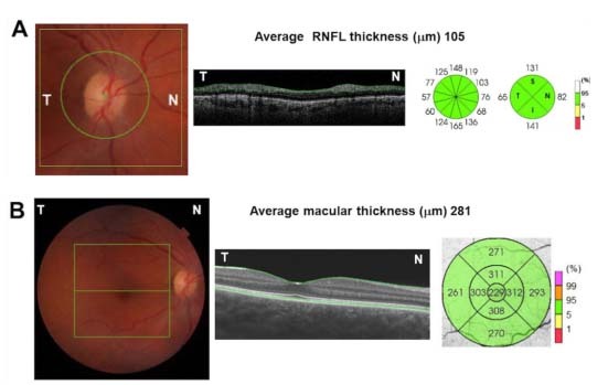
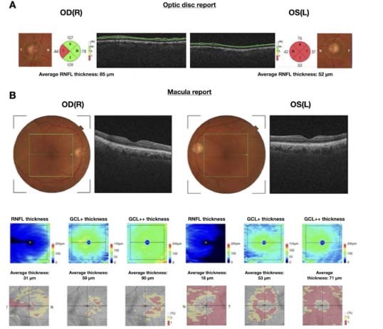
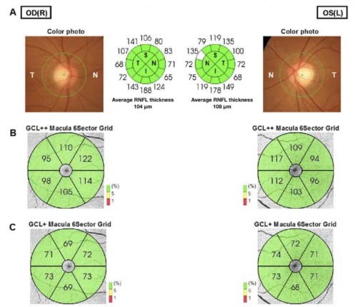
Structural imaging of the brain is the most widely used diagnostic tool for investigating neurodegenerative diseases. More advanced structural imaging techniques have been applied to early or prodromic phases, but they are expensive and not widely available. Therefore, it is highly desirable to search for noninvasive, easily accessible, low-cost clinical biomarkers suitable for large-scale population screening, in order to focus on making diagnoses at the earliest stages of the disease. In this scenario, imaging studies focusing on the structures of the retina have increasingly been used for evaluating neurodegenerative diseases. The retina shares embryological, histological, biochemical, microvascular and neurotransmitter similarities with the cerebral cortex, thus making it a uniquely promising biomarker for neurodegenerative diseases. Optical coherence tomography is a modern noninvasive imaging technique that provides high-resolution two-dimensional cross-sectional images and quantitative reproducible three-dimensional volumetric measurements of the optic nerve head and retina. This technology is widely used in ophthalmology practice for diagnosing and following up several eye diseases, such as glaucoma, diabetic retinopathy and age-related macular degeneration. Its clinical impact on neurodegenerative diseases has raised enormous interest over recent years, as several clinical studies have demonstrated that these diseases give rise to reduced thickness of the inner retinal nerve fiber layer, mainly composed of retinal ganglion cells and their axons. In this review, we aimed to address the clinical utility of optical coherence tomography for diagnosing and evaluating different neurodegenerative diseases, to show the potential of this noninvasive and easily accessible method.
脑结构影像学是目前用于研究神经退行性疾病的最广泛应用的诊断工具。更先进的结构成像技术已被应用于早期或前驱阶段,但它们昂贵且尚未广泛应用。因此,非常需要寻找非侵入性、易于获得、低成本的临床生物标志物,以适合大规模人群筛查,从而专注于在疾病的最早阶段进行诊断。在这种情况下,越来越多的关注视网膜结构的影像学研究被用于评估神经退行性疾病。视网膜与大脑皮层在胚胎发生、组织学、生物化学、微血管和神经递质方面具有相似性,因此成为神经退行性疾病极具前景的生物标志物。光学相干断层扫描是一种现代的非侵入性成像技术,可提供视神经头和视网膜的高分辨率二维横截面图像和可重复的定量三维容积测量。该技术在眼科实践中广泛用于诊断和随访多种眼病,如青光眼、糖尿病视网膜病变和年龄相关性黄斑变性。近年来,其在神经退行性疾病中的临床应用引起了极大的兴趣,因为几项临床研究表明,这些疾病会导致视网膜神经纤维层内层厚度变薄,主要由视网膜神经节细胞及其轴突组成。在这篇综述中,我们旨在探讨光学相干断层扫描在诊断和评估不同神经退行性疾病中的临床应用,展示这种非侵入性、易于获得的方法的潜力。