• 文献检索
  • 文档翻译
  • 深度研究
  • 学术资讯
  • Suppr Zotero 插件Zotero 插件
  • 邀请有礼
  • 套餐&价格
  • 历史记录
应用&插件
Suppr Zotero 插件Zotero 插件浏览器插件Mac 客户端Windows 客户端微信小程序
定价
高级版会员购买积分包购买API积分包
服务
文献检索文档翻译深度研究API 文档MCP 服务
关于我们
关于 Suppr公司介绍联系我们用户协议隐私条款
关注我们

Suppr 超能文献

核心技术专利:CN118964589B侵权必究
粤ICP备2023148730 号-1Suppr @ 2026

文献检索

告别复杂PubMed语法,用中文像聊天一样搜索,搜遍4000万医学文献。AI智能推荐,让科研检索更轻松。

立即免费搜索

文件翻译

保留排版,准确专业,支持PDF/Word/PPT等文件格式,支持 12+语言互译。

免费翻译文档

深度研究

AI帮你快速写综述,25分钟生成高质量综述,智能提取关键信息,辅助科研写作。

立即免费体验

腹膜透析流出液中细胞外囊泡的表征及蛋白质组学分析

Characterization and proteomic profile of extracellular vesicles from peritoneal dialysis efflux.

作者信息

Carreras-Planella Laura, Soler-Majoral Jordi, Rubio-Esteve Cristina, Lozano-Ramos Sara Inés, Franquesa Marcella, Bonet Josep, Troya-Saborido Maria Isabel, Borràs Francesc Enric

机构信息

REMAR-IVECAT Group, "Germans Trias i Pujol" Health Science Research Institute, Can Ruti Campus, Badalona, Spain.

Department of Cell Biology, Physiology and Immunology, Autonomous University of Barcelona, Barcelona, Spain.

出版信息

PLoS One. 2017 May 10;12(5):e0176987. doi: 10.1371/journal.pone.0176987. eCollection 2017.

DOI:10.1371/journal.pone.0176987
PMID:28489901
原文链接:https://pmc.ncbi.nlm.nih.gov/articles/PMC5425196/
Abstract

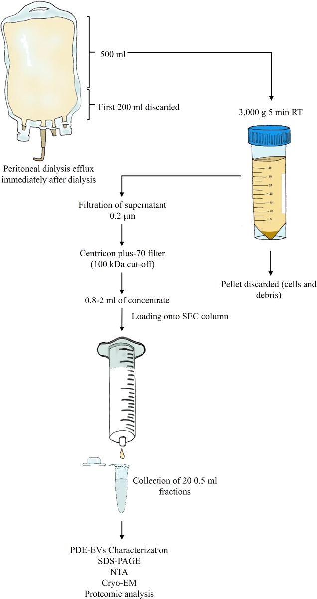
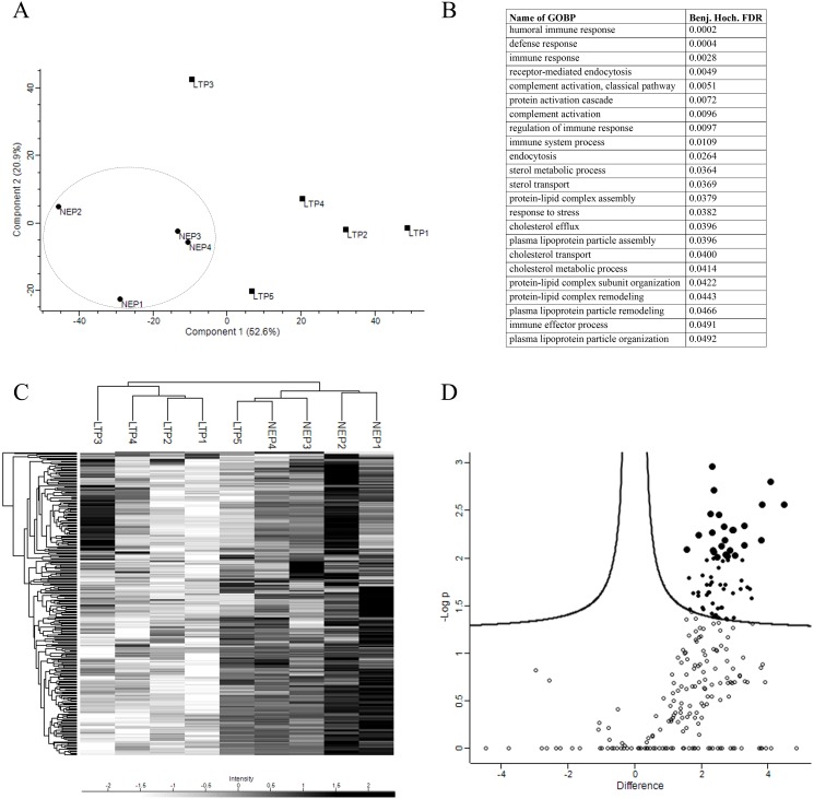
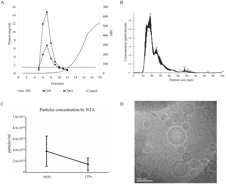
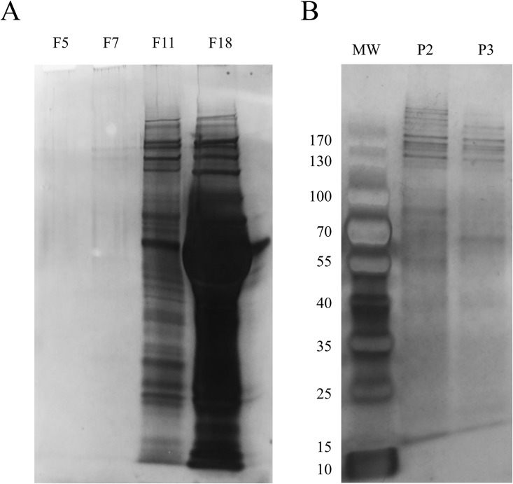
Peritoneal Dialysis (PD) is considered the best option for a cost-effective mid-term dialysis in patients with Chronic Renal Failure. However, functional failure of the peritoneal membrane (PM) force many patients to stop PD treatment and start haemodialysis. Currently, PM functionality is monitored by the peritoneal equilibration test, a tedious technique that often show changes when the membrane damage is advanced. As in other pathologies, the identification and characterization of extracellular vesicles (EVs) in the peritoneal dialysis efflux (PDE) may represent a non-invasive alternative to identify biomarkers of membrane failure. Using size-exclusion chromatography, we isolated EVs from PDE in a group of patients. Vesicles were characterized by the presence of tetraspanin markers, nanoparticle tracking analysis profile, cryo-electron microscopy and mass spectrometry. Here, we report the isolation and characterization of PDE-EVs. Based on mass spectrometry, we have found a set of well-conserved proteins among patients. Interestingly, the peptide profile also revealed remarkable changes between newly enrolled and longer-treated PD patients. These results are the first step to the identification of PDE-EVs based new markers of PM damage, which could support clinicians in their decision-making in a non-invasive manner.

摘要

腹膜透析(PD)被认为是慢性肾衰竭患者进行具有成本效益的中期透析的最佳选择。然而,腹膜(PM)功能衰竭迫使许多患者停止PD治疗并开始血液透析。目前,PM功能通过腹膜平衡试验进行监测,这是一种繁琐的技术,当膜损伤进展时常常显示出变化。与其他病症一样,腹膜透析流出液(PDE)中细胞外囊泡(EVs)的鉴定和表征可能代表一种识别膜衰竭生物标志物的非侵入性替代方法。我们使用尺寸排阻色谱法从一组患者的PDE中分离出EVs。通过四跨膜蛋白标记物的存在、纳米颗粒跟踪分析图谱、冷冻电子显微镜和质谱对囊泡进行表征。在此,我们报告PDE-EVs的分离和表征。基于质谱分析,我们在患者中发现了一组保守性良好的蛋白质。有趣的是,肽谱还揭示了新入组和长期接受PD治疗的患者之间存在显著差异。这些结果是鉴定基于PDE-EVs的PM损伤新标志物的第一步,这可以以非侵入性方式支持临床医生进行决策。

https://cdn.ncbi.nlm.nih.gov/pmc/blobs/8579/5425196/373917ac9e6f/pone.0176987.g006.jpg
https://cdn.ncbi.nlm.nih.gov/pmc/blobs/8579/5425196/e87d9378957a/pone.0176987.g001.jpg
https://cdn.ncbi.nlm.nih.gov/pmc/blobs/8579/5425196/346f15e0bb90/pone.0176987.g002.jpg
https://cdn.ncbi.nlm.nih.gov/pmc/blobs/8579/5425196/df3789e7d3b2/pone.0176987.g003.jpg
https://cdn.ncbi.nlm.nih.gov/pmc/blobs/8579/5425196/350333c58ea6/pone.0176987.g004.jpg
https://cdn.ncbi.nlm.nih.gov/pmc/blobs/8579/5425196/1ae7067330a2/pone.0176987.g005.jpg
https://cdn.ncbi.nlm.nih.gov/pmc/blobs/8579/5425196/373917ac9e6f/pone.0176987.g006.jpg

相似文献

1
Characterization and proteomic profile of extracellular vesicles from peritoneal dialysis efflux.腹膜透析流出液中细胞外囊泡的表征及蛋白质组学分析
PLoS One. 2017 May 10;12(5):e0176987. doi: 10.1371/journal.pone.0176987. eCollection 2017.
2
Proteomic profiling of peritoneal dialysis effluent-derived extracellular vesicles: a longitudinal study.腹透液来源细胞外囊泡的蛋白质组学分析:一项纵向研究。
J Nephrol. 2019 Dec;32(6):1021-1031. doi: 10.1007/s40620-019-00658-3. Epub 2019 Oct 15.
3
Multiple extracellular vesicle types in peritoneal dialysis effluent are prominent and contain known biomarkers.腹膜透析流出液中的多种细胞外囊泡类型显著,且含有已知的生物标志物。
PLoS One. 2017 Jun 8;12(6):e0178601. doi: 10.1371/journal.pone.0178601. eCollection 2017.
4
Glycoprotein 96 in Peritoneal Dialysis Effluent-Derived Extracellular Vesicles: A Tool for Evaluating Peritoneal Transport Properties and Inflammatory Status.腹透液来源外泌体中的糖蛋白 96:评估腹膜转运特性和炎症状态的工具。
Front Immunol. 2022 Feb 10;13:824278. doi: 10.3389/fimmu.2022.824278. eCollection 2022.
5
Proteomic Characterization of Peritoneal Extracellular Vesicles in a Mouse Model of Peritoneal Fibrosis.腹膜纤维化小鼠模型中腹膜细胞外囊泡的蛋白质组学特征
J Proteome Res. 2023 Mar 3;22(3):908-918. doi: 10.1021/acs.jproteome.2c00713. Epub 2023 Jan 17.
6
A cost-effective method to get insight into the peritoneal dialysate effluent proteome.一种了解腹膜透析液蛋白质组的经济有效方法。
J Proteomics. 2016 Aug 11;145:207-213. doi: 10.1016/j.jprot.2016.05.010. Epub 2016 May 20.
7
Combinatorial Peptide Ligand Library and two dimensional electrophoresis: new frontiers in the study of peritoneal dialysis effluent in pediatric patients.组合肽配体文库与二维电泳:小儿患者腹膜透析流出液研究的新前沿
J Proteomics. 2015 Feb 26;116:68-80. doi: 10.1016/j.jprot.2015.01.003. Epub 2015 Jan 10.
8
Proteomic analysis of extracellular vesicles secreted by primary human epithelial endometrial cells reveals key proteins related to embryo implantation.人原发性上皮子宫内膜细胞分泌的细胞外囊泡的蛋白质组学分析揭示了与胚胎植入相关的关键蛋白。
Reprod Biol Endocrinol. 2022 Jan 3;20(1):3. doi: 10.1186/s12958-021-00879-x.
9
A prospective, proteomics study identified potential biomarkers of encapsulating peritoneal sclerosis in peritoneal effluent.一项前瞻性的蛋白质组学研究鉴定出腹水中包裹性腹膜硬化症的潜在生物标志物。
Kidney Int. 2017 Oct;92(4):988-1002. doi: 10.1016/j.kint.2017.03.030. Epub 2017 Jul 1.
10
Proteomic Research in Peritoneal Dialysis.腹膜透析中的蛋白质组学研究。
Int J Mol Sci. 2020 Jul 31;21(15):5489. doi: 10.3390/ijms21155489.

引用本文的文献

1
Development of a Sampling and Storage Protocol of Extracellular Vesicles (EVs)-Establishment of the First EV Biobank for Polytraumatized Patients.建立创伤患者的首个细胞外囊泡(EV)生物样本库:一种 EV 采样和存储方案的开发。
Int J Mol Sci. 2024 May 22;25(11):5645. doi: 10.3390/ijms25115645.
2
Extracellular Vesicles of Patients on Peritoneal Dialysis Inhibit the TGF-β- and PDGF-B-Mediated Fibrotic Processes.腹透患者的细胞外囊泡抑制 TGF-β 和 PDGF-B 介导的纤维化过程。
Cells. 2024 Mar 29;13(7):605. doi: 10.3390/cells13070605.
3
Differentially Expressed microRNAs in Peritoneal Dialysis Effluent-Derived Exosomes from the Patients with Ultrafiltration Failure.

本文引用的文献

1
Size-Exclusion Chromatography-based isolation minimally alters Extracellular Vesicles' characteristics compared to precipitating agents.基于尺寸排阻色谱法的分离与沉淀剂相比,最小程度地改变了细胞外囊泡的特征。
Sci Rep. 2016 Sep 19;6:33641. doi: 10.1038/srep33641.
2
The Perseus computational platform for comprehensive analysis of (prote)omics data.Perseus 计算平台,用于全面分析(蛋白质组学)数据。
Nat Methods. 2016 Sep;13(9):731-40. doi: 10.1038/nmeth.3901. Epub 2016 Jun 27.
3
Evidence-Based Clinical Use of Nanoscale Extracellular Vesicles in Nanomedicine.
超滤衰竭患者腹腔透析流出液衍生外泌体中的差异表达 microRNAs。
Genet Res (Camb). 2022 Aug 31;2022:2276175. doi: 10.1155/2022/2276175. eCollection 2022.
4
Proteomics and Extracellular Vesicles as Novel Biomarker Sources in Peritoneal Dialysis in Children.蛋白质组学和细胞外囊泡作为儿童腹膜透析中新型生物标志物来源。
Int J Mol Sci. 2022 May 18;23(10):5655. doi: 10.3390/ijms23105655.
5
Glycoprotein 96 in Peritoneal Dialysis Effluent-Derived Extracellular Vesicles: A Tool for Evaluating Peritoneal Transport Properties and Inflammatory Status.腹透液来源外泌体中的糖蛋白 96:评估腹膜转运特性和炎症状态的工具。
Front Immunol. 2022 Feb 10;13:824278. doi: 10.3389/fimmu.2022.824278. eCollection 2022.
6
The Past, the Present, and the Future of the Size Exclusion Chromatography in Extracellular Vesicles Separation.大小排阻色谱法在细胞外囊泡分离中的过去、现在和未来。
Viruses. 2021 Nov 13;13(11):2272. doi: 10.3390/v13112272.
7
Extracellular Vesicles and Their Relationship with the Heart-Kidney Axis, Uremia and Peritoneal Dialysis.细胞外囊泡及其与心肾轴、尿毒症和腹膜透析的关系。
Toxins (Basel). 2021 Nov 4;13(11):778. doi: 10.3390/toxins13110778.
8
Extracellular vesicles as regulators of kidney function and disease.细胞外囊泡作为肾功能和疾病的调节因子
Intensive Care Med Exp. 2020 Dec 18;8(Suppl 1):22. doi: 10.1186/s40635-020-00306-2.
9
Exosomes in Nephropathies: A Rich Source of Novel Biomarkers.肾病中的外泌体:新型生物标志物的丰富来源。
Dis Markers. 2020 Aug 12;2020:8897833. doi: 10.1155/2020/8897833. eCollection 2020.
10
Proteomic Research in Peritoneal Dialysis.腹膜透析中的蛋白质组学研究。
Int J Mol Sci. 2020 Jul 31;21(15):5489. doi: 10.3390/ijms21155489.
纳米医学中基于证据的纳米级细胞外囊泡的临床应用。
ACS Nano. 2016 Apr 26;10(4):3886-99. doi: 10.1021/acsnano.5b08015. Epub 2016 Mar 15.
4
Applying extracellular vesicles based therapeutics in clinical trials - an ISEV position paper.应用细胞外囊泡为基础的治疗方法于临床试验——ISEV 立场文件。
J Extracell Vesicles. 2015 Dec 31;4:30087. doi: 10.3402/jev.v4.30087. eCollection 2015.
5
Hemodialysis versus peritoneal dialysis, which is cost-effective?血液透析与腹膜透析,哪种更具成本效益?
Saudi J Kidney Dis Transpl. 2015 Sep;26(5):962-5. doi: 10.4103/1319-2442.164578.
6
Extracellular Vesicles in Renal Diseases: More than Novel Biomarkers?肾脏疾病中的细胞外囊泡:不止是新型生物标志物?
J Am Soc Nephrol. 2016 Jan;27(1):12-26. doi: 10.1681/ASN.2015010074. Epub 2015 Aug 6.
7
Size-exclusion chromatography-based enrichment of extracellular vesicles from urine samples.基于排阻色谱法从尿液样本中富集细胞外囊泡。
J Extracell Vesicles. 2015 May 28;4:27369. doi: 10.3402/jev.v4.27369. eCollection 2015.
8
InteractiVenn: a web-based tool for the analysis of sets through Venn diagrams.InteractiVenn:一款基于网络的通过维恩图进行集合分析的工具。
BMC Bioinformatics. 2015 May 22;16(1):169. doi: 10.1186/s12859-015-0611-3.
9
Biological properties of extracellular vesicles and their physiological functions.细胞外囊泡的生物学特性及其生理功能。
J Extracell Vesicles. 2015 May 14;4:27066. doi: 10.3402/jev.v4.27066. eCollection 2015.
10
A microrna screen to identify regulators of peritoneal fibrosis in a rat model of peritoneal dialysis.一项用于在腹膜透析大鼠模型中鉴定腹膜纤维化调节因子的微小RNA筛选。
BMC Nephrol. 2015 Apr 9;16:48. doi: 10.1186/s12882-015-0039-z.