• 文献检索
  • 文档翻译
  • 深度研究
  • 学术资讯
  • Suppr Zotero 插件Zotero 插件
  • 邀请有礼
  • 套餐&价格
  • 历史记录
应用&插件
Suppr Zotero 插件Zotero 插件浏览器插件Mac 客户端Windows 客户端微信小程序
定价
高级版会员购买积分包购买API积分包
服务
文献检索文档翻译深度研究API 文档MCP 服务
关于我们
关于 Suppr公司介绍联系我们用户协议隐私条款
关注我们

Suppr 超能文献

核心技术专利:CN118964589B侵权必究
粤ICP备2023148730 号-1Suppr @ 2026

文献检索

告别复杂PubMed语法,用中文像聊天一样搜索,搜遍4000万医学文献。AI智能推荐,让科研检索更轻松。

立即免费搜索

文件翻译

保留排版,准确专业,支持PDF/Word/PPT等文件格式,支持 12+语言互译。

免费翻译文档

深度研究

AI帮你快速写综述,25分钟生成高质量综述,智能提取关键信息,辅助科研写作。

立即免费体验

脾脏巨噬细胞的募集会导致血管紧张素 II 诱导的心肌纤维化和高血压。

Recruitment of macrophages from the spleen contributes to myocardial fibrosis and hypertension induced by angiotensin II.

作者信息

Wang Ning-Ping, Erskine James, Zhang Wei-Wei, Zheng Rong-Hua, Zhang Li-Hui, Duron Garret, Gendreau Julian, Zhao Zhi-Qing

机构信息

1 Cardiovascular Research Laboratory, Mercer University School of Medicine, USA.

2 Department of Internal Medicine, Navicent Health, USA.

出版信息

J Renin Angiotensin Aldosterone Syst. 2017 Apr-Jun;18(2):1470320317706653. doi: 10.1177/1470320317706653.

DOI:10.1177/1470320317706653
PMID:28490219
原文链接:https://pmc.ncbi.nlm.nih.gov/articles/PMC5843916/
Abstract

INTRODUCTION

The purpose of this study was to determine whether macrophages migrated from the spleen are associated with angiotensin II-induced cardiac fibrosis and hypertension.

METHODS

Sprague-Dawley rats were subjected to angiotensin II infusion in vehicle (500 ng/kg/min) for up to four weeks. In splenectomy, the spleen was removed before angiotensin II infusion. In the angiotensin II AT1 receptor blockade, telmisartan was administered by gastric gavage (10 mg/kg/day) during angiotensin II infusion. The heart and aorta were isolated for Western blot analysis and immunohistochemistry.

RESULTS

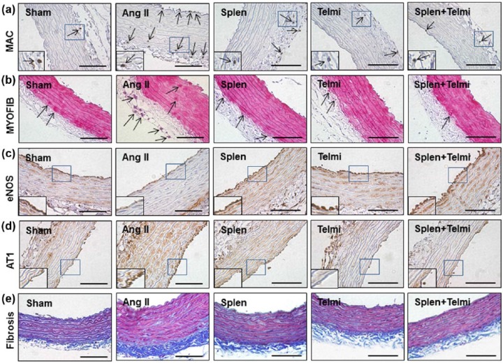
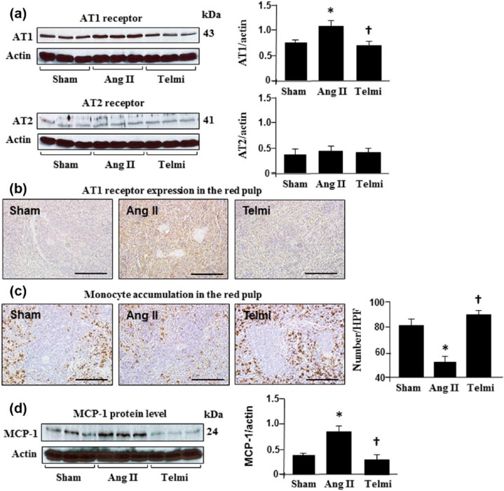
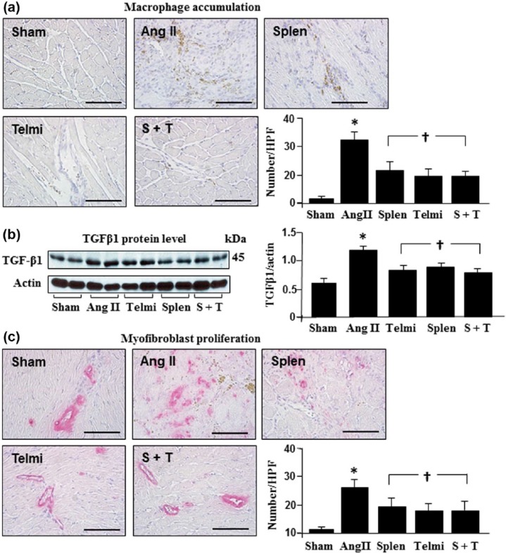
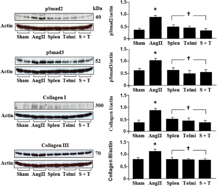
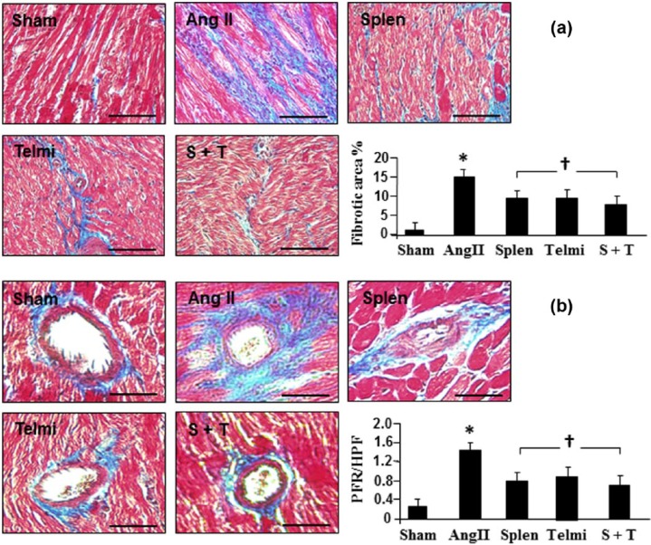
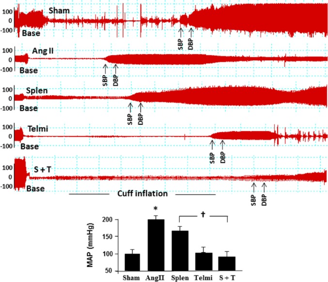
Angiotensin II infusion caused a significant reduction in the number of monocytes in the spleen through the AT1 receptor-activated monocyte chemoattractant protein-1. Comparison of angiotensin II infusion, splenectomy and telmisartan comparatively reduced the recruitment of macrophages into the heart. Associated with this change, transforming growth factor β1 expression and myofibroblast proliferation were inhibited, and Smad2/3 and collagen I/III were downregulated. Furthermore, interstitial/perivascular fibrosis was attenuated. These modifications occurred in coincidence with reduced blood pressure. At week 4, invasion of macrophages and myofibroblasts in the thoracic aorta was attenuated and expression of endothelial nitric oxide synthase was upregulated, along with a reduction in aortic fibrosis.

CONCLUSIONS

These results suggest that macrophages when recruited into the heart and aorta from the spleen potentially contribute to angiotensin II-induced cardiac fibrosis and hypertension.

摘要

引言

本研究的目的是确定从脾脏迁移的巨噬细胞是否与血管紧张素II诱导的心脏纤维化和高血压有关。

方法

将Sprague-Dawley大鼠用载体(500 ng/kg/分钟)输注血管紧张素II,持续长达四周。在脾切除术中,在输注血管紧张素II之前切除脾脏。在血管紧张素II AT1受体阻断中,在输注血管紧张素II期间通过灌胃给予替米沙坦(10 mg/kg/天)。分离心脏和主动脉用于蛋白质印迹分析和免疫组织化学。

结果

血管紧张素II输注通过AT1受体激活的单核细胞趋化蛋白-1导致脾脏中单核细胞数量显著减少。血管紧张素II输注、脾切除术和替米沙坦的比较相对减少了巨噬细胞向心脏的募集。与此变化相关,转化生长因子β1表达和肌成纤维细胞增殖受到抑制,Smad2/3和胶原蛋白I/III下调。此外,间质/血管周围纤维化减轻。这些改变与血压降低同时发生。在第4周,胸主动脉中巨噬细胞和成纤维细胞的浸润减弱,内皮型一氧化氮合酶的表达上调,同时主动脉纤维化减少。

结论

这些结果表明,从脾脏募集到心脏和主动脉中的巨噬细胞可能促成血管紧张素II诱导的心脏纤维化和高血压。

https://cdn.ncbi.nlm.nih.gov/pmc/blobs/52e5/5843916/d8124bda84ea/10.1177_1470320317706653-fig7.jpg
https://cdn.ncbi.nlm.nih.gov/pmc/blobs/52e5/5843916/e931db12df66/10.1177_1470320317706653-fig1.jpg
https://cdn.ncbi.nlm.nih.gov/pmc/blobs/52e5/5843916/e724bfb8b726/10.1177_1470320317706653-fig2.jpg
https://cdn.ncbi.nlm.nih.gov/pmc/blobs/52e5/5843916/60f70374a568/10.1177_1470320317706653-fig3.jpg
https://cdn.ncbi.nlm.nih.gov/pmc/blobs/52e5/5843916/5cbe2c7421b0/10.1177_1470320317706653-fig4.jpg
https://cdn.ncbi.nlm.nih.gov/pmc/blobs/52e5/5843916/d876ac61c712/10.1177_1470320317706653-fig5.jpg
https://cdn.ncbi.nlm.nih.gov/pmc/blobs/52e5/5843916/0c4dc562e782/10.1177_1470320317706653-fig6.jpg
https://cdn.ncbi.nlm.nih.gov/pmc/blobs/52e5/5843916/d8124bda84ea/10.1177_1470320317706653-fig7.jpg

相似文献

1
Recruitment of macrophages from the spleen contributes to myocardial fibrosis and hypertension induced by angiotensin II.脾脏巨噬细胞的募集会导致血管紧张素 II 诱导的心肌纤维化和高血压。
J Renin Angiotensin Aldosterone Syst. 2017 Apr-Jun;18(2):1470320317706653. doi: 10.1177/1470320317706653.
2
Edaravone inhibits pressure overload-induced cardiac fibrosis and dysfunction by reducing expression of angiotensin II AT1 receptor.依达拉奉通过降低血管紧张素II 1型受体的表达来抑制压力超负荷诱导的心脏纤维化和功能障碍。
Drug Des Devel Ther. 2017 Oct 16;11:3019-3033. doi: 10.2147/DDDT.S144807. eCollection 2017.
3
Preservation of Glucagon-Like Peptide-1 Level Attenuates Angiotensin II-Induced Tissue Fibrosis by Altering AT1/AT 2 Receptor Expression and Angiotensin-Converting Enzyme 2 Activity in Rat Heart.通过改变大鼠心脏中AT1/AT2受体表达和血管紧张素转换酶2活性来维持胰高血糖素样肽-1水平可减轻血管紧张素II诱导的组织纤维化。
Cardiovasc Drugs Ther. 2015 Jun;29(3):243-55. doi: 10.1007/s10557-015-6592-7.
4
Angiotensin II AT1 receptor alters ACE2 activity, eNOS expression and CD44-hyaluronan interaction in rats with hypertension and myocardial fibrosis.血管紧张素II 1型受体改变高血压和心肌纤维化大鼠的血管紧张素转换酶2活性、内皮型一氧化氮合酶表达及CD44与透明质酸的相互作用。
Life Sci. 2016 May 15;153:141-52. doi: 10.1016/j.lfs.2016.04.013. Epub 2016 Apr 13.
5
Steroidogenic acute regulatory protein/aldosterone synthase mediates angiotensin II-induced cardiac fibrosis and hypertrophy.类固醇生成急性调节蛋白/醛固酮合酶介导血管紧张素 II 诱导的心脏纤维化和肥大。
Mol Biol Rep. 2020 Feb;47(2):1207-1222. doi: 10.1007/s11033-019-05222-7. Epub 2019 Dec 9.
6
Postconditioning attenuates coronary perivascular and interstitial fibrosis through modulating angiotensin II receptors and angiotensin-converting enzyme 2 after myocardial infarction.心肌梗死后,后适应通过调节血管紧张素II受体和血管紧张素转换酶2减轻冠状动脉血管周围和间质纤维化。
J Surg Res. 2017 May 1;211:178-190. doi: 10.1016/j.jss.2016.11.046. Epub 2016 Dec 5.
7
Attenuation of myocardial fibrosis with curcumin is mediated by modulating expression of angiotensin II AT1/AT2 receptors and ACE2 in rats.姜黄素对大鼠心肌纤维化的减轻作用是通过调节血管紧张素II AT1/AT2受体和ACE2的表达来介导的。
Drug Des Devel Ther. 2015 Nov 11;9:6043-54. doi: 10.2147/DDDT.S95333. eCollection 2015.
8
C-reactive protein promotes cardiac fibrosis and inflammation in angiotensin II-induced hypertensive cardiac disease.C 反应蛋白促进血管紧张素Ⅱ诱导的高血压性心脏病中的心肌纤维化和炎症。
Hypertension. 2010 Apr;55(4):953-60. doi: 10.1161/HYPERTENSIONAHA.109.140608. Epub 2010 Feb 15.
9
Liraglutide attenuates cardiac remodeling and improves heart function after abdominal aortic constriction through blocking angiotensin II type 1 receptor in rats.利拉鲁肽通过阻断大鼠血管紧张素II 1型受体减轻腹主动脉缩窄后的心脏重塑并改善心脏功能。
Drug Des Devel Ther. 2019 Aug 6;13:2745-2757. doi: 10.2147/DDDT.S213910. eCollection 2019.
10
Candesartan ameliorates arsenic-induced hypertensive vascular remodeling by regularizing angiotensin II and TGF-beta signaling in rats.坎地沙坦通过调节大鼠体内的血管紧张素II和转化生长因子-β信号通路,改善砷诱导的高血压血管重塑。
Toxicology. 2016 Dec 30;374:29-41. doi: 10.1016/j.tox.2016.11.015. Epub 2016 Nov 23.

引用本文的文献

1
Pathophysiology of Angiotensin II-Mediated Hypertension, Cardiac Hypertrophy, and Failure: A Perspective from Macrophages.血管紧张素II介导的高血压、心脏肥大和心力衰竭的病理生理学:来自巨噬细胞的视角
Cells. 2024 Dec 4;13(23):2001. doi: 10.3390/cells13232001.
2
Heart Failure with Preserved Ejection Fraction and Cardiac Amyloidosis in the Aging Heart.衰老心脏中的射血分数保留型心力衰竭和心脏淀粉样变性。
Int J Mol Sci. 2024 Oct 26;25(21):11519. doi: 10.3390/ijms252111519.
3
Isoliensinine Attenuates Renal Fibrosis and Inhibits TGF-β1/Smad2/3 Signaling Pathway in Spontaneously Hypertensive Rats.

本文引用的文献

1
Role of the Renin-Angiotensin-Aldosterone System beyond Blood Pressure Regulation: Molecular and Cellular Mechanisms Involved in End-Organ Damage during Arterial Hypertension.肾素-血管紧张素-醛固酮系统在血压调节之外的作用:动脉高血压期间终末器官损伤所涉及的分子和细胞机制
Int J Mol Sci. 2016 Jun 23;17(7):797. doi: 10.3390/ijms17070797.
2
Angiotensin II AT1 receptor alters ACE2 activity, eNOS expression and CD44-hyaluronan interaction in rats with hypertension and myocardial fibrosis.血管紧张素II 1型受体改变高血压和心肌纤维化大鼠的血管紧张素转换酶2活性、内皮型一氧化氮合酶表达及CD44与透明质酸的相互作用。
Life Sci. 2016 May 15;153:141-52. doi: 10.1016/j.lfs.2016.04.013. Epub 2016 Apr 13.
3
异莲心碱减轻自发性高血压大鼠的肾纤维化并抑制 TGF-β1/Smad2/3 信号通路。
Drug Des Devel Ther. 2023 Sep 7;17:2749-2762. doi: 10.2147/DDDT.S414179. eCollection 2023.
4
Cardiovascular Protection with a Long-Acting GLP-1 Receptor Agonist Liraglutide: An Experimental Update.长效 GLP-1 受体激动剂利拉鲁肽的心血管保护作用:实验更新。
Molecules. 2023 Feb 1;28(3):1369. doi: 10.3390/molecules28031369.
5
Identification of Key Genes from the Visceral Adipose Tissues of Overweight/Obese Adults with Hypertension through Transcriptome Sequencing.通过转录组测序从超重/肥胖高血压成年患者的内脏脂肪组织中鉴定关键基因
Cytogenet Genome Res. 2022;162(10):541-559. doi: 10.1159/000528702. Epub 2022 Dec 15.
6
The E3 ubiquitin ligase WWP2 regulates pro-fibrogenic monocyte infiltration and activity in heart fibrosis.E3 泛素连接酶 WWP2 调节心肌纤维化中促纤维化单核细胞浸润和活性。
Nat Commun. 2022 Nov 30;13(1):7375. doi: 10.1038/s41467-022-34971-6.
7
Organized Chaos: Deciphering Immune Cell Heterogeneity's Role in Inflammation in the Heart.组织混乱:解析免疫细胞异质性在心脏炎症中的作用。
Biomolecules. 2021 Dec 22;12(1):11. doi: 10.3390/biom12010011.
8
Alteration of circadian machinery in monocytes underlies chronic kidney disease-associated cardiac inflammation and fibrosis.昼夜节律机制在单核细胞中的改变是慢性肾脏病相关心脏炎症和纤维化的基础。
Nat Commun. 2021 May 13;12(1):2783. doi: 10.1038/s41467-021-23050-x.
9
[Angiotensin Ⅱ inhibits AMPK/SIRT1 pathway by inducing oxidative stress in RAW264.7 macrophages].[血管紧张素Ⅱ通过诱导RAW264.7巨噬细胞氧化应激抑制AMPK/SIRT1通路]
Nan Fang Yi Ke Da Xue Xue Bao. 2021 Mar 25;41(3):384-390. doi: 10.12122/j.issn.1673-4254.2021.03.10.
10
Nlrp3 Deficiency Alleviates Angiotensin II-Induced Cardiomyopathy by Inhibiting Mitochondrial Dysfunction.Nlrp3 缺失通过抑制线粒体功能障碍缓解血管紧张素 II 诱导的心肌病。
Oxid Med Cell Longev. 2021 Feb 5;2021:6679100. doi: 10.1155/2021/6679100. eCollection 2021.
ACE inhibition, ACE2 and angiotensin-(1-7) axis in kidney and cardiac inflammation and fibrosis.
肾脏和心脏炎症及纤维化中的血管紧张素转换酶抑制、血管紧张素转换酶2与血管紧张素-(1-7)轴
Pharmacol Res. 2016 May;107:154-162. doi: 10.1016/j.phrs.2016.03.018. Epub 2016 Mar 17.
4
Macrophage depletion lowers blood pressure and restores sympathetic nerve α2-adrenergic receptor function in mesenteric arteries of DOCA-salt hypertensive rats.巨噬细胞耗竭可降低去氧皮质酮盐(DOCA)-盐高血压大鼠肠系膜动脉的血压,并恢复交感神经α2-肾上腺素能受体功能。
Am J Physiol Heart Circ Physiol. 2015 Oct;309(7):H1186-97. doi: 10.1152/ajpheart.00283.2015. Epub 2015 Aug 28.
5
Involvement of IGF-1 and Akt in M1/M2 activation state in bone marrow-derived macrophages.胰岛素样生长因子-1(IGF-1)和蛋白激酶B(Akt)参与骨髓源性巨噬细胞的M1/M2活化状态。
Exp Cell Res. 2015 Jul 15;335(2):258-68. doi: 10.1016/j.yexcr.2015.05.015. Epub 2015 May 28.
6
Preservation of Glucagon-Like Peptide-1 Level Attenuates Angiotensin II-Induced Tissue Fibrosis by Altering AT1/AT 2 Receptor Expression and Angiotensin-Converting Enzyme 2 Activity in Rat Heart.通过改变大鼠心脏中AT1/AT2受体表达和血管紧张素转换酶2活性来维持胰高血糖素样肽-1水平可减轻血管紧张素II诱导的组织纤维化。
Cardiovasc Drugs Ther. 2015 Jun;29(3):243-55. doi: 10.1007/s10557-015-6592-7.
7
Novel mechanism of cardiac protection by valsartan: synergetic roles of TGF-β1 and HIF-1α in Ang II-mediated fibrosis after myocardial infarction.缬沙坦心脏保护的新机制:转化生长因子-β1和低氧诱导因子-1α在心肌梗死后血管紧张素II介导的纤维化中的协同作用
J Cell Mol Med. 2015 Aug;19(8):1773-82. doi: 10.1111/jcmm.12551. Epub 2015 Mar 30.
8
Monocytes in myocardial infarction.心肌梗死中的单核细胞。
Arterioscler Thromb Vasc Biol. 2015 May;35(5):1066-70. doi: 10.1161/ATVBAHA.114.304652. Epub 2015 Mar 19.
9
Activated macrophages as a feeder layer for growth of resident cardiac progenitor cells.活化巨噬细胞作为心脏常驻祖细胞生长的饲养层。
Cytotechnology. 2016 Aug;68(4):665-74. doi: 10.1007/s10616-014-9814-0. Epub 2014 Nov 29.
10
Angiotensin II-induced endothelial dysfunction is temporally linked with increases in interleukin-6 and vascular macrophage accumulation.血管紧张素 II 诱导的内皮功能障碍与白细胞介素-6 的增加以及血管巨噬细胞积聚在时间上相关联。
Front Physiol. 2014 Oct 29;5:396. doi: 10.3389/fphys.2014.00396. eCollection 2014.