Section on Model Synaptic Systems, Laboratory of Molecular Physiology, NIAAA, NIH, Bethesda, Maryland, 20892, USA.
Department of Psychiatry, Columbia University, and Division of Integrative Neuroscience, New York State Psychiatric Institute, New York, NY, 10032, USA.
Sci Rep. 2017 May 10;7(1):1674. doi: 10.1038/s41598-017-01900-3.
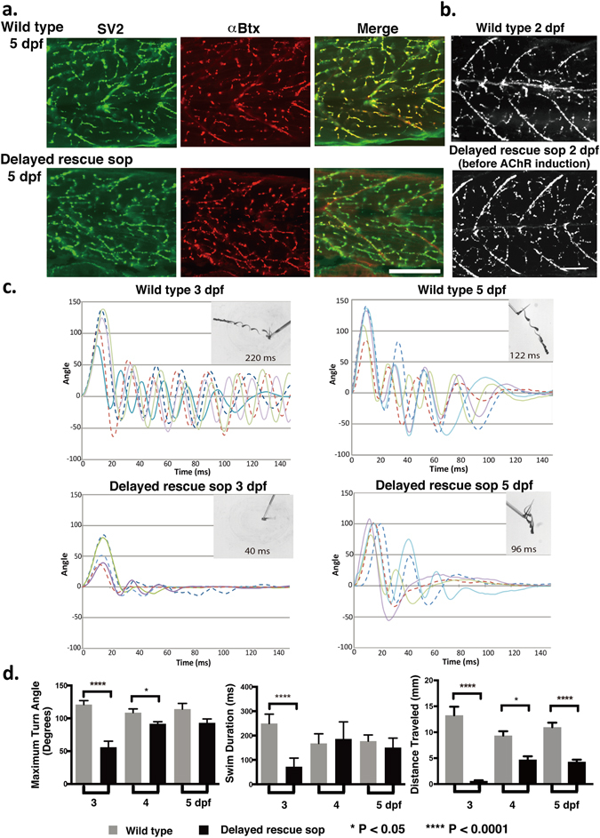
The formation and function of synapses are tightly orchestrated by the precise timing of expression of specific molecules during development. In this study, we determined how manipulating the timing of expression of postsynaptic acetylcholine receptors (AChRs) impacts presynaptic release by establishing a genetically engineered zebrafish line in which we can freely control the timing of AChR expression in an AChR-less fish background. With the delayed induction of AChR expression after an extensive period of AChR-less development, paralyzed fish displayed a remarkable level of recovery, exhibiting a robust escape response following developmental delay. Despite their apparent behavioral rescue, synapse formation in these fish was significantly altered as a result of delayed AChR expression. Motor neuron innervation determined the sites for AChR clustering, a complete reversal of normal neuromuscular junction (NMJ) development where AChR clustering precedes innervation. Most importantly, among the three modes of presynaptic vesicle release, only the spontaneous release machinery was strongly suppressed in these fish, while evoked vesicle release remained relatively unaffected. Such a specific presynaptic change, which may constitute a part of the compensatory mechanism in response to the absence of postsynaptic AChRs, may underlie symptoms of neuromuscular diseases characterized by reduced AChRs, such as myasthenia gravis.
突触的形成和功能是通过在发育过程中特定分子的精确表达时间来紧密协调的。在这项研究中,我们通过建立一个可在无乙酰胆碱受体(AChR)的鱼类背景中自由控制 AChR 表达时间的基因工程斑马鱼系,确定了改变突触后 AChR 表达时间如何影响突触前释放。通过在无 AChR 发育的很长一段时间后延迟 AChR 表达的诱导,瘫痪的鱼表现出了显著的恢复水平,在发育延迟后表现出了强大的逃避反应。尽管这些鱼的行为明显得到了挽救,但由于 AChR 表达的延迟,它们的突触形成发生了显著的改变。运动神经元的神经支配决定了 AChR 聚集的部位,这完全颠覆了正常的神经肌肉接头(NMJ)发育,其中 AChR 聚集先于神经支配。最重要的是,在三种类型的突触前囊泡释放中,只有自发释放机制在这些鱼中受到强烈抑制,而诱发的囊泡释放相对不受影响。这种特定的突触前变化可能构成了对缺乏突触后 AChR 反应的一部分补偿机制,可能是由 AChR 减少引起的神经肌肉疾病的症状的基础,如重症肌无力。