Xin Long, Xu Weixing, Yu Leijun, Fan Shunwu, Wang Wei, Yu Fang, Wang Zhenbin
Department of Spine Surgery, Tongde Hospital of Zhejiang Province, Hangzhou, China.
Department of Orthopedics, the Affiliated Sir Run Run Shaw Hospital, Zhejiang University, Zhejiang, China.
J Orthop Surg Res. 2017 May 12;12(1):73. doi: 10.1186/s13018-017-0572-5.
Growth of nerve fibers has been shown to occur in a rabbit model of intravertebral disc degeneration (IVD) induced by needle puncture. As nerve growth may underlie the process of chronic pain in humans affected by disc degeneration, we sought to investigate the factors underlying nerve ingrowth in a minimally invasive annulotomy rabbit model of IVD by comparing the effects of empty disc defects with those of defects filled with poly(lactic-co-glycolic acid)/fibrin gel (PLGA) plugs.
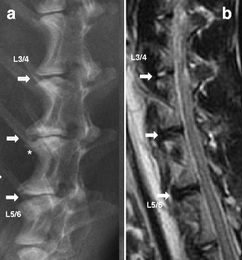
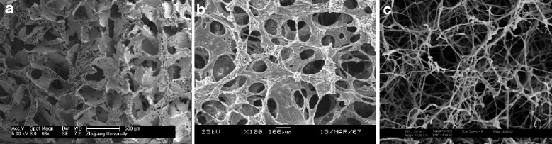
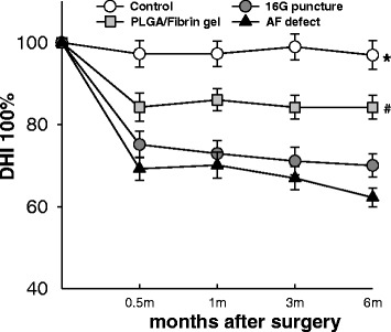
New Zealand white rabbits (n = 24) received annular injuries at three lumbar levels (L3/4, L4/5, and L5/6). The discs were randomly assigned to four groups: (a) annular defect (1.8-mm diameter; 4-mm depth) by mini-trephine, (b) annular defect implanted with a PLGA scaffold containing a fibrin gel, (c) annular puncture by a 16G needle (5-mm depth), and (d) uninjured L2/3 disc (control). Disc degeneration was evaluated by radiography, MRI, histology, real-time PCR, and analysis of proteoglycan (PG) content. Nerve ingrowth into the discs was assessed by immunostaining with the nerve marker protein gene product 9.5.
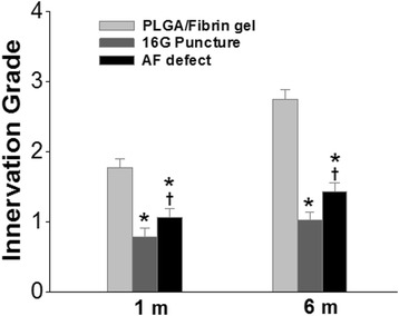
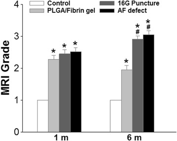
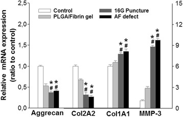
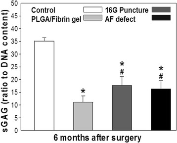
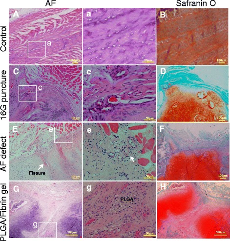
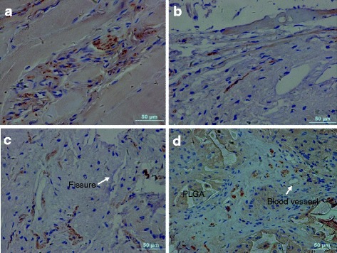
Injured discs showed a progressive disc space narrowing with significant disc degeneration and proteoglycan loss, as confirmed by imaging results, molecular and compositional analysis, and histological examinations. In 16G punctured discs, nerve ingrowth was observed on the surface of scar tissue. In annular defects, nerve fibers were found to be distributed along small fissures within the fibrocartilaginous-like tissue that filled the AF. In discs filled with PLGA/ fibrin gel, more nerve fibers were observed growing deeper into the inner AF along the open annular track. In addition, innervations scores showed significantly higher than those of punctured discs and empty defects. A limited vascular proliferation was found in the injured sites and regenerated tissues.
Nerve ingrowth was significantly higher in PLGA/fibrin-filled discs than in empty defects. Possible explanations include (i) annular fissures along the defect and early loss of proteoglycan may facilitate the ingrowth process and (ii) biodegradable PLGA/fibrin gel may promote adverse growth of nerves and blood vessels into deeper parts of injured disc. The rabbit annular defect model of disc degeneration appears suitable to investigate the effects of nerve ingrowth in relation to pain generation.
在针刺诱导的兔椎间盘退变(IVD)模型中已证实有神经纤维生长。由于神经生长可能是人类椎间盘退变所致慢性疼痛过程的基础,我们试图通过比较空椎间盘缺损与填充聚乳酸-乙醇酸共聚物/纤维蛋白凝胶(PLGA)栓子的缺损对神经长入的影响,来研究微创环形切开兔IVD模型中神经长入的相关因素。
24只新西兰白兔在三个腰椎节段(L3/4、L4/5和L5/6)接受环形损伤。椎间盘被随机分为四组:(a)用微型环锯制作的环形缺损(直径1.8mm;深度4mm),(b)植入含有纤维蛋白凝胶的PLGA支架的环形缺损,(c)用16G针进行的环形穿刺(深度5mm),以及(d)未受伤的L2/3椎间盘(对照)。通过X线摄影、MRI、组织学、实时PCR和蛋白聚糖(PG)含量分析来评估椎间盘退变。用神经标志物蛋白基因产物9.5进行免疫染色来评估神经向椎间盘内的长入情况。
影像学结果、分子和成分分析以及组织学检查证实,受伤的椎间盘显示出椎间盘间隙逐渐变窄,伴有明显的椎间盘退变和蛋白聚糖丢失。在16G穿刺的椎间盘中,在瘢痕组织表面观察到神经长入。在环形缺损中,发现神经纤维沿填充纤维环的类纤维软骨样组织内的小裂隙分布。在填充PLGA/纤维蛋白凝胶的椎间盘中,观察到更多神经纤维沿开放的环形通道深入到纤维环内层生长。此外,神经支配评分显示明显高于穿刺椎间盘和空缺损组。在损伤部位和再生组织中发现有限的血管增生。
填充PLGA/纤维蛋白的椎间盘中神经长入明显高于空缺损组。可能的解释包括:(i)沿缺损的环形裂隙和蛋白聚糖的早期丢失可能促进了长入过程;(ii)可生物降解的PLGA/纤维蛋白凝胶可能促进神经和血管向受伤椎间盘深部的不良生长。兔椎间盘退变环形缺损模型似乎适合研究神经长入与疼痛产生的关系。